Magnesium »
PDB 5m5l-5mh5 »
5maq »
Magnesium in PDB 5maq: Crystal Structure of Polyphosphate Kinase From Meiothermus Ruber Bound to Adp and Ppi
Protein crystallography data
The structure of Crystal Structure of Polyphosphate Kinase From Meiothermus Ruber Bound to Adp and Ppi, PDB code: 5maq
was solved by
S.Gerhardt,
O.Einsle,
F.Kemper,
N.Schwarzer,
with X-Ray Crystallography technique. A brief refinement statistics is given in the table below:
| Resolution Low / High (Å) | 117.49 / 2.46 |
| Space group | P 43 21 2 |
| Cell size a, b, c (Å), α, β, γ (°) | 166.159, 166.159, 94.985, 90.00, 90.00, 90.00 |
| R / Rfree (%) | 19.1 / 23.4 |
Magnesium Binding Sites:
The binding sites of Magnesium atom in the Crystal Structure of Polyphosphate Kinase From Meiothermus Ruber Bound to Adp and Ppi
(pdb code 5maq). This binding sites where shown within
5.0 Angstroms radius around Magnesium atom.
In total 4 binding sites of Magnesium where determined in the Crystal Structure of Polyphosphate Kinase From Meiothermus Ruber Bound to Adp and Ppi, PDB code: 5maq:
Jump to Magnesium binding site number: 1; 2; 3; 4;
In total 4 binding sites of Magnesium where determined in the Crystal Structure of Polyphosphate Kinase From Meiothermus Ruber Bound to Adp and Ppi, PDB code: 5maq:
Jump to Magnesium binding site number: 1; 2; 3; 4;
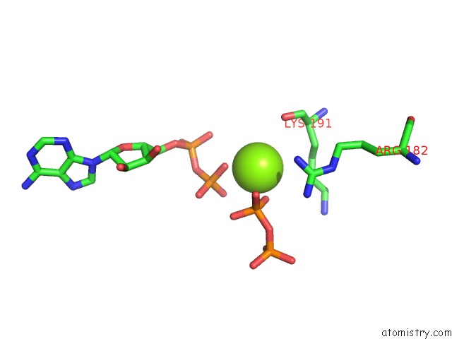
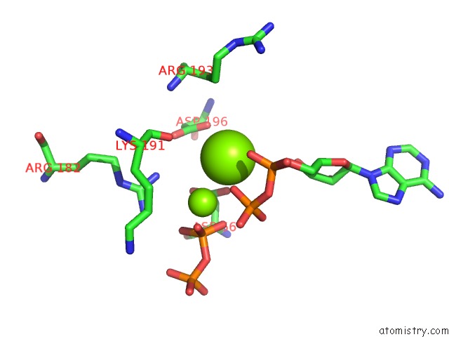
Magnesium binding site 1 out of 4 in 5maq
Go back to
Magnesium binding site 1 out
of 4 in the Crystal Structure of Polyphosphate Kinase From Meiothermus Ruber Bound to Adp and Ppi
Mono view
Stereo pair view
Mono view
Stereo pair view
A full contact list of Magnesium with other atoms in the Mg binding
site number 1 of Crystal Structure of Polyphosphate Kinase From Meiothermus Ruber Bound to Adp and Ppi within 5.0Å range:
|
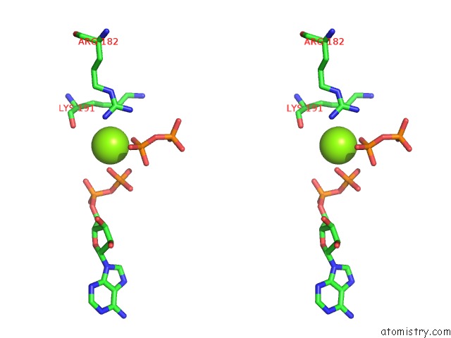
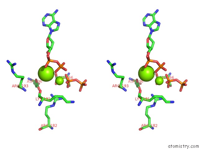
Magnesium binding site 2 out of 4 in 5maq
Go back to
Magnesium binding site 2 out
of 4 in the Crystal Structure of Polyphosphate Kinase From Meiothermus Ruber Bound to Adp and Ppi
Mono view
Stereo pair view
Mono view
Stereo pair view
A full contact list of Magnesium with other atoms in the Mg binding
site number 2 of Crystal Structure of Polyphosphate Kinase From Meiothermus Ruber Bound to Adp and Ppi within 5.0Å range:
|
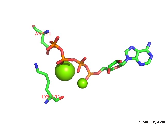
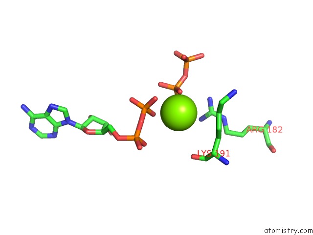
Magnesium binding site 3 out of 4 in 5maq
Go back to
Magnesium binding site 3 out
of 4 in the Crystal Structure of Polyphosphate Kinase From Meiothermus Ruber Bound to Adp and Ppi
Mono view
Stereo pair view
Mono view
Stereo pair view
A full contact list of Magnesium with other atoms in the Mg binding
site number 3 of Crystal Structure of Polyphosphate Kinase From Meiothermus Ruber Bound to Adp and Ppi within 5.0Å range:
|
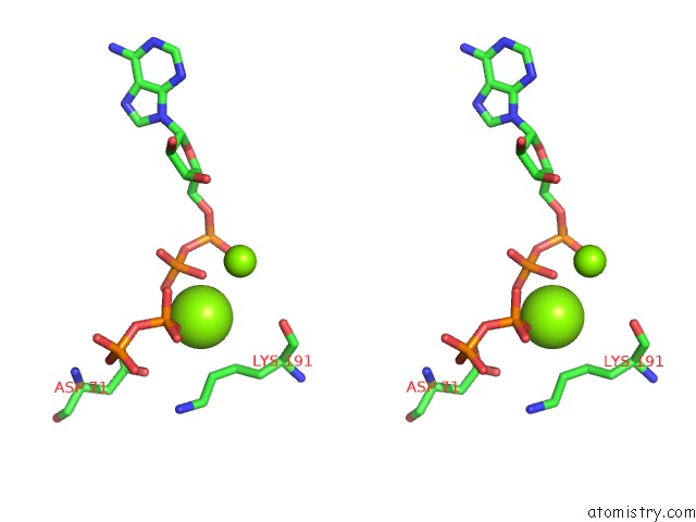
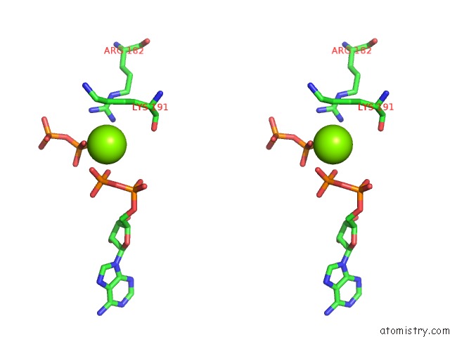
Magnesium binding site 4 out of 4 in 5maq
Go back to
Magnesium binding site 4 out
of 4 in the Crystal Structure of Polyphosphate Kinase From Meiothermus Ruber Bound to Adp and Ppi
Mono view
Stereo pair view
Mono view
Stereo pair view
A full contact list of Magnesium with other atoms in the Mg binding
site number 4 of Crystal Structure of Polyphosphate Kinase From Meiothermus Ruber Bound to Adp and Ppi within 5.0Å range:
|
Reference:
A.E.Parnell,
S.Mordhorst,
F.Kemper,
M.Giurrandino,
J.P.Prince,
N.J.Schwarzer,
A.Hofer,
D.Wohlwend,
H.J.Jessen,
S.Gerhardt,
O.Einsle,
P.C.F.Oyston,
J.N.Andexer,
P.L.Roach.
Substrate Recognition and Mechanism Revealed By Ligand-Bound Polyphosphate Kinase 2 Structures. Proc. Natl. Acad. Sci. V. 115 3350 2018U.S.A..
ISSN: ESSN 1091-6490
PubMed: 29531036
DOI: 10.1073/PNAS.1710741115
Page generated: Sun Sep 29 21:17:40 2024
ISSN: ESSN 1091-6490
PubMed: 29531036
DOI: 10.1073/PNAS.1710741115
Last articles
F in 4JLNF in 4JLT
F in 4JLM
F in 4JLG
F in 4JJU
F in 4JLK
F in 4JKV
F in 4JLJ
F in 4JJS
F in 4JII