Magnesium »
PDB 5m5l-5mh5 »
5mb9 »
Magnesium in PDB 5mb9: Crystal Structure of the Eukaryotic Ribosome Associated Complex (Rac), A Unique HSP70/HSP40 Pair
Protein crystallography data
The structure of Crystal Structure of the Eukaryotic Ribosome Associated Complex (Rac), A Unique HSP70/HSP40 Pair, PDB code: 5mb9
was solved by
A.Gumiero,
F.A.Weyer,
G.Valentin Gese,
K.Lapouge,
I.Sinning,
with X-Ray Crystallography technique. A brief refinement statistics is given in the table below:
| Resolution Low / High (Å) | 47.97 / 3.20 |
| Space group | C 2 2 21 |
| Cell size a, b, c (Å), α, β, γ (°) | 93.918, 179.359, 155.455, 90.00, 90.00, 90.00 |
| R / Rfree (%) | 22.7 / 28.5 |
Magnesium Binding Sites:
The binding sites of Magnesium atom in the Crystal Structure of the Eukaryotic Ribosome Associated Complex (Rac), A Unique HSP70/HSP40 Pair
(pdb code 5mb9). This binding sites where shown within
5.0 Angstroms radius around Magnesium atom.
In total 2 binding sites of Magnesium where determined in the Crystal Structure of the Eukaryotic Ribosome Associated Complex (Rac), A Unique HSP70/HSP40 Pair, PDB code: 5mb9:
Jump to Magnesium binding site number: 1; 2;
In total 2 binding sites of Magnesium where determined in the Crystal Structure of the Eukaryotic Ribosome Associated Complex (Rac), A Unique HSP70/HSP40 Pair, PDB code: 5mb9:
Jump to Magnesium binding site number: 1; 2;
Magnesium binding site 1 out of 2 in 5mb9
Go back to
Magnesium binding site 1 out
of 2 in the Crystal Structure of the Eukaryotic Ribosome Associated Complex (Rac), A Unique HSP70/HSP40 Pair
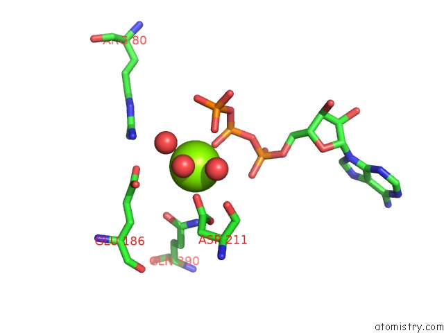
Mono view
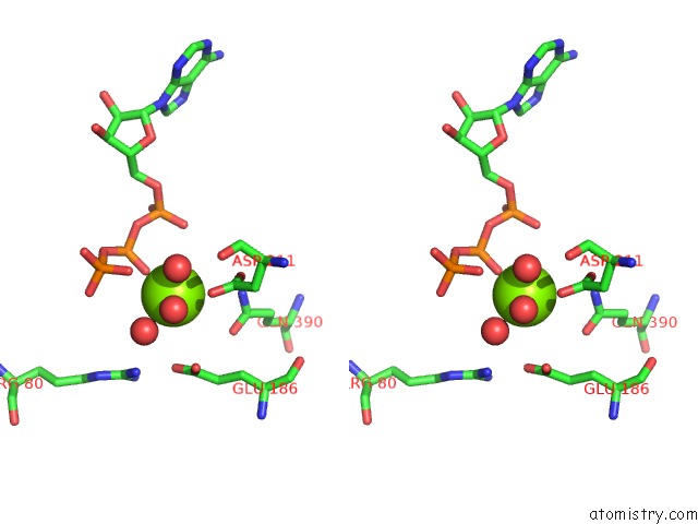
Stereo pair view
Mono view
Stereo pair view
A full contact list of Magnesium with other atoms in the Mg binding
site number 1 of Crystal Structure of the Eukaryotic Ribosome Associated Complex (Rac), A Unique HSP70/HSP40 Pair within 5.0Å range:
|
Magnesium binding site 2 out of 2 in 5mb9
Go back to
Magnesium binding site 2 out
of 2 in the Crystal Structure of the Eukaryotic Ribosome Associated Complex (Rac), A Unique HSP70/HSP40 Pair
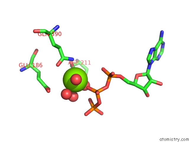
Mono view
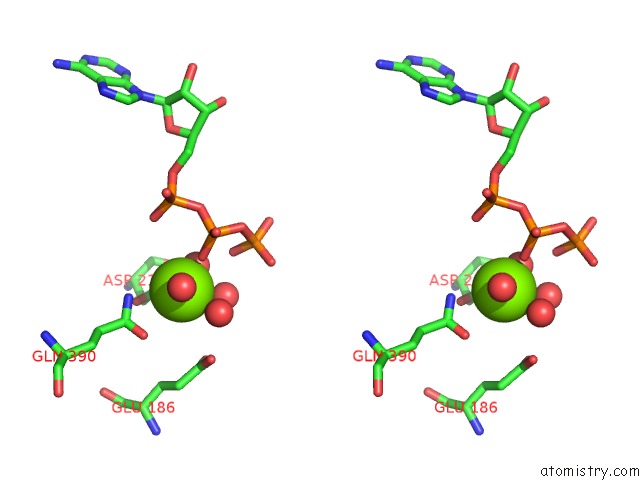
Stereo pair view
Mono view
Stereo pair view
A full contact list of Magnesium with other atoms in the Mg binding
site number 2 of Crystal Structure of the Eukaryotic Ribosome Associated Complex (Rac), A Unique HSP70/HSP40 Pair within 5.0Å range:
|
Reference:
F.A.Weyer,
A.Gumiero,
G.V.Gese,
K.Lapouge,
I.Sinning.
Structural Insights Into A Unique HSP70-HSP40 Interaction in the Eukaryotic Ribosome-Associated Complex. Nat. Struct. Mol. Biol. V. 24 144 2017.
ISSN: ESSN 1545-9985
PubMed: 28067917
DOI: 10.1038/NSMB.3349
Page generated: Sun Sep 29 21:18:11 2024
ISSN: ESSN 1545-9985
PubMed: 28067917
DOI: 10.1038/NSMB.3349
Last articles
Ca in 5SZLCa in 5SY1
Ca in 5SWI
Ca in 5SVE
Ca in 5SSX
Ca in 5SV0
Ca in 5STD
Ca in 5SSZ
Ca in 5SSY
Ca in 5SIC