Magnesium »
PDB 5m5i-5mh1 »
5mga »
Magnesium in PDB 5mga: Structure of the CPF1 Endonuclease R-Loop Complex After Dna Cleavage
Protein crystallography data
The structure of Structure of the CPF1 Endonuclease R-Loop Complex After Dna Cleavage, PDB code: 5mga
was solved by
G.Montoya,
S.Stella,
with X-Ray Crystallography technique. A brief refinement statistics is given in the table below:
| Resolution Low / High (Å) | 39.58 / 3.00 |
| Space group | C 2 2 21 |
| Cell size a, b, c (Å), α, β, γ (°) | 85.223, 137.652, 320.513, 90.00, 90.00, 90.00 |
| R / Rfree (%) | 24.2 / 26.5 |
Magnesium Binding Sites:
The binding sites of Magnesium atom in the Structure of the CPF1 Endonuclease R-Loop Complex After Dna Cleavage
(pdb code 5mga). This binding sites where shown within
5.0 Angstroms radius around Magnesium atom.
In total 2 binding sites of Magnesium where determined in the Structure of the CPF1 Endonuclease R-Loop Complex After Dna Cleavage, PDB code: 5mga:
Jump to Magnesium binding site number: 1; 2;
In total 2 binding sites of Magnesium where determined in the Structure of the CPF1 Endonuclease R-Loop Complex After Dna Cleavage, PDB code: 5mga:
Jump to Magnesium binding site number: 1; 2;
Magnesium binding site 1 out of 2 in 5mga
Go back to
Magnesium binding site 1 out
of 2 in the Structure of the CPF1 Endonuclease R-Loop Complex After Dna Cleavage
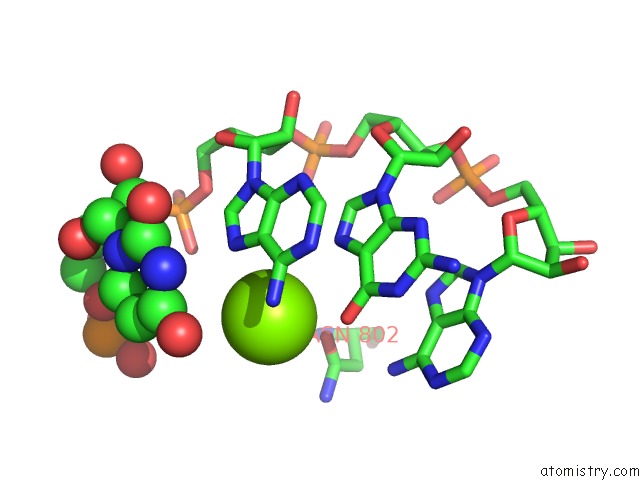
Mono view
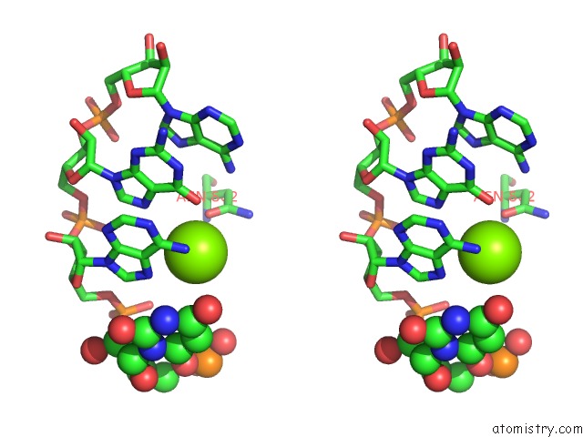
Stereo pair view
Mono view
Stereo pair view
A full contact list of Magnesium with other atoms in the Mg binding
site number 1 of Structure of the CPF1 Endonuclease R-Loop Complex After Dna Cleavage within 5.0Å range:
|
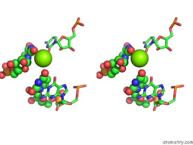
Magnesium binding site 2 out of 2 in 5mga
Go back to
Magnesium binding site 2 out
of 2 in the Structure of the CPF1 Endonuclease R-Loop Complex After Dna Cleavage
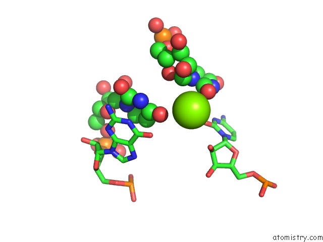
Mono view
Stereo pair view
Mono view
Stereo pair view
A full contact list of Magnesium with other atoms in the Mg binding
site number 2 of Structure of the CPF1 Endonuclease R-Loop Complex After Dna Cleavage within 5.0Å range:
|
Reference:
S.Stella,
P.Alcon,
G.Montoya.
Structure of the CPF1 Endonuclease R-Loop Complex After Target Dna Cleavage. Nature V. 546 559 2017.
ISSN: ISSN 0028-0836
PubMed: 28562584
DOI: 10.1038/NATURE22398
Page generated: Sun Sep 29 21:22:00 2024
ISSN: ISSN 0028-0836
PubMed: 28562584
DOI: 10.1038/NATURE22398
Last articles
Zn in 9JYWZn in 9IR4
Zn in 9IR3
Zn in 9GMX
Zn in 9GMW
Zn in 9JEJ
Zn in 9ERF
Zn in 9ERE
Zn in 9EGV
Zn in 9EGW