Magnesium »
PDB 5mh6-5mtv »
5mtv »
Magnesium in PDB 5mtv: Active Structure of EHD4 Complexed with Atp-Gamma-S
Protein crystallography data
The structure of Active Structure of EHD4 Complexed with Atp-Gamma-S, PDB code: 5mtv
was solved by
A.A.Melo,
O.Daumke,
with X-Ray Crystallography technique. A brief refinement statistics is given in the table below:
| Resolution Low / High (Å) | 48.50 / 2.79 |
| Space group | P 42 21 2 |
| Cell size a, b, c (Å), α, β, γ (°) | 199.973, 199.973, 41.537, 90.00, 90.00, 90.00 |
| R / Rfree (%) | 22.7 / 24.3 |
Magnesium Binding Sites:
The binding sites of Magnesium atom in the Active Structure of EHD4 Complexed with Atp-Gamma-S
(pdb code 5mtv). This binding sites where shown within
5.0 Angstroms radius around Magnesium atom.
In total only one binding site of Magnesium was determined in the Active Structure of EHD4 Complexed with Atp-Gamma-S, PDB code: 5mtv:
In total only one binding site of Magnesium was determined in the Active Structure of EHD4 Complexed with Atp-Gamma-S, PDB code: 5mtv:
Magnesium binding site 1 out of 1 in 5mtv
Go back to
Magnesium binding site 1 out
of 1 in the Active Structure of EHD4 Complexed with Atp-Gamma-S
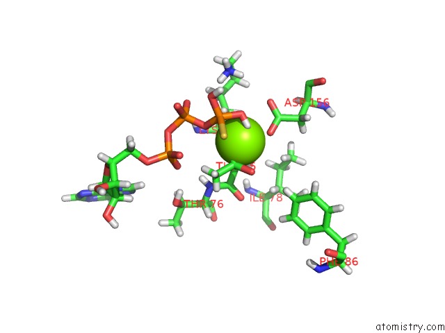
Mono view
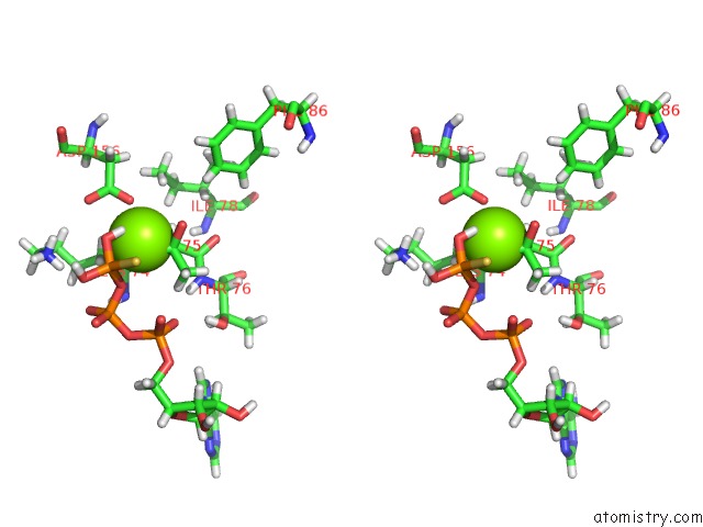
Stereo pair view
Mono view
Stereo pair view
A full contact list of Magnesium with other atoms in the Mg binding
site number 1 of Active Structure of EHD4 Complexed with Atp-Gamma-S within 5.0Å range:
|
Reference:
A.A.Melo,
B.G.Hegde,
C.Shah,
E.Larsson,
J.M.Isas,
S.Kunz,
R.Lundmark,
R.Langen,
O.Daumke.
Structural Insights Into the Activation Mechanism of Dynamin-Like Ehd Atpases. Proc. Natl. Acad. Sci. V. 114 5629 2017U.S.A..
ISSN: ESSN 1091-6490
PubMed: 28228524
DOI: 10.1073/PNAS.1614075114
Page generated: Tue Aug 12 15:32:16 2025
ISSN: ESSN 1091-6490
PubMed: 28228524
DOI: 10.1073/PNAS.1614075114
Last articles
Mg in 5SI5Mg in 5SI4
Mg in 5SI3
Mg in 5SI2
Mg in 5SI0
Mg in 5SHZ
Mg in 5SI1
Mg in 5SHX
Mg in 5SHW
Mg in 5SHY