Magnesium »
PDB 5mtv-5n77 »
5n2q »
Magnesium in PDB 5n2q: Mobm Relaxase Domain (Mobv; MOB_PRE) Bound to 26NT PMV158 Orit Dna
Protein crystallography data
The structure of Mobm Relaxase Domain (Mobv; MOB_PRE) Bound to 26NT PMV158 Orit Dna, PDB code: 5n2q
was solved by
S.Russi,
D.R.Boer,
M.Coll,
with X-Ray Crystallography technique. A brief refinement statistics is given in the table below:
| Resolution Low / High (Å) | 55.73 / 2.00 |
| Space group | P 1 21 1 |
| Cell size a, b, c (Å), α, β, γ (°) | 43.576, 52.853, 56.035, 90.00, 95.88, 90.00 |
| R / Rfree (%) | 16.6 / 23.8 |
Other elements in 5n2q:
The structure of Mobm Relaxase Domain (Mobv; MOB_PRE) Bound to 26NT PMV158 Orit Dna also contains other interesting chemical elements:
| Chlorine | (Cl) | 1 atom |
| Sodium | (Na) | 1 atom |
Magnesium Binding Sites:
The binding sites of Magnesium atom in the Mobm Relaxase Domain (Mobv; MOB_PRE) Bound to 26NT PMV158 Orit Dna
(pdb code 5n2q). This binding sites where shown within
5.0 Angstroms radius around Magnesium atom.
In total only one binding site of Magnesium was determined in the Mobm Relaxase Domain (Mobv; MOB_PRE) Bound to 26NT PMV158 Orit Dna, PDB code: 5n2q:
In total only one binding site of Magnesium was determined in the Mobm Relaxase Domain (Mobv; MOB_PRE) Bound to 26NT PMV158 Orit Dna, PDB code: 5n2q:
Magnesium binding site 1 out of 1 in 5n2q
Go back to
Magnesium binding site 1 out
of 1 in the Mobm Relaxase Domain (Mobv; MOB_PRE) Bound to 26NT PMV158 Orit Dna
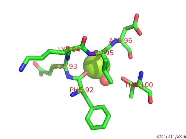
Mono view
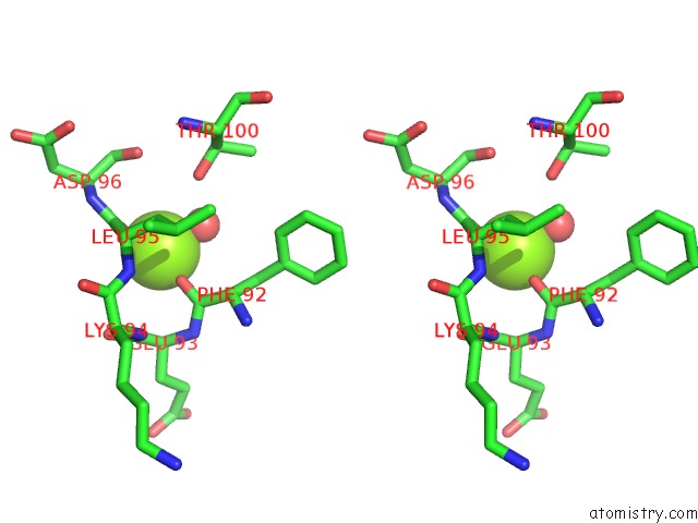
Stereo pair view
Mono view
Stereo pair view
A full contact list of Magnesium with other atoms in the Mg binding
site number 1 of Mobm Relaxase Domain (Mobv; MOB_PRE) Bound to 26NT PMV158 Orit Dna within 5.0Å range:
|
Reference:
R.Pluta,
D.R.Boer,
F.Lorenzo-Diaz,
S.Russi,
H.Gomez,
C.Fernandez-Lopez,
R.Perez-Luque,
M.Orozco,
M.Espinosa,
M.Coll.
Structural Basis of A Histidine-Dna Nicking/Joining Mechanism For Gene Transfer and Promiscuous Spread of Antibiotic Resistance. Proc. Natl. Acad. Sci. V. 114 E6526 2017U.S.A..
ISSN: ESSN 1091-6490
PubMed: 28739894
DOI: 10.1073/PNAS.1702971114
Page generated: Sun Sep 29 23:04:16 2024
ISSN: ESSN 1091-6490
PubMed: 28739894
DOI: 10.1073/PNAS.1702971114
Last articles
Zn in 9J0NZn in 9J0O
Zn in 9J0P
Zn in 9FJX
Zn in 9EKB
Zn in 9C0F
Zn in 9CAH
Zn in 9CH0
Zn in 9CH3
Zn in 9CH1