Magnesium »
PDB 5mtv-5n77 »
5n77 »
Magnesium in PDB 5n77: Crystal Structure of the Cytosolic Domain of the Cora Magnesium Channel From Escherichia Coli in Complex with Magnesium
Protein crystallography data
The structure of Crystal Structure of the Cytosolic Domain of the Cora Magnesium Channel From Escherichia Coli in Complex with Magnesium, PDB code: 5n77
was solved by
M.Lerche,
H.Sandhu,
L.Flockner,
M.Hogbom,
M.Rapp,
with X-Ray Crystallography technique. A brief refinement statistics is given in the table below:
| Resolution Low / High (Å) | 29.84 / 2.80 |
| Space group | P 1 21 1 |
| Cell size a, b, c (Å), α, β, γ (°) | 73.313, 117.453, 91.114, 90.00, 103.27, 90.00 |
| R / Rfree (%) | 19.5 / 23.6 |
Magnesium Binding Sites:
The binding sites of Magnesium atom in the Crystal Structure of the Cytosolic Domain of the Cora Magnesium Channel From Escherichia Coli in Complex with Magnesium
(pdb code 5n77). This binding sites where shown within
5.0 Angstroms radius around Magnesium atom.
In total 7 binding sites of Magnesium where determined in the Crystal Structure of the Cytosolic Domain of the Cora Magnesium Channel From Escherichia Coli in Complex with Magnesium, PDB code: 5n77:
Jump to Magnesium binding site number: 1; 2; 3; 4; 5; 6; 7;
In total 7 binding sites of Magnesium where determined in the Crystal Structure of the Cytosolic Domain of the Cora Magnesium Channel From Escherichia Coli in Complex with Magnesium, PDB code: 5n77:
Jump to Magnesium binding site number: 1; 2; 3; 4; 5; 6; 7;
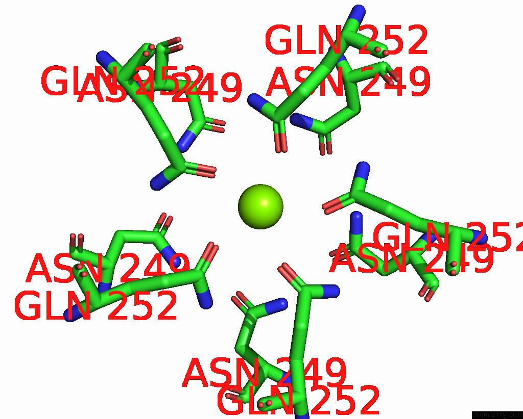
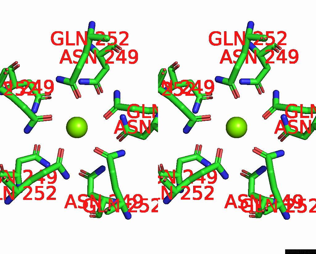
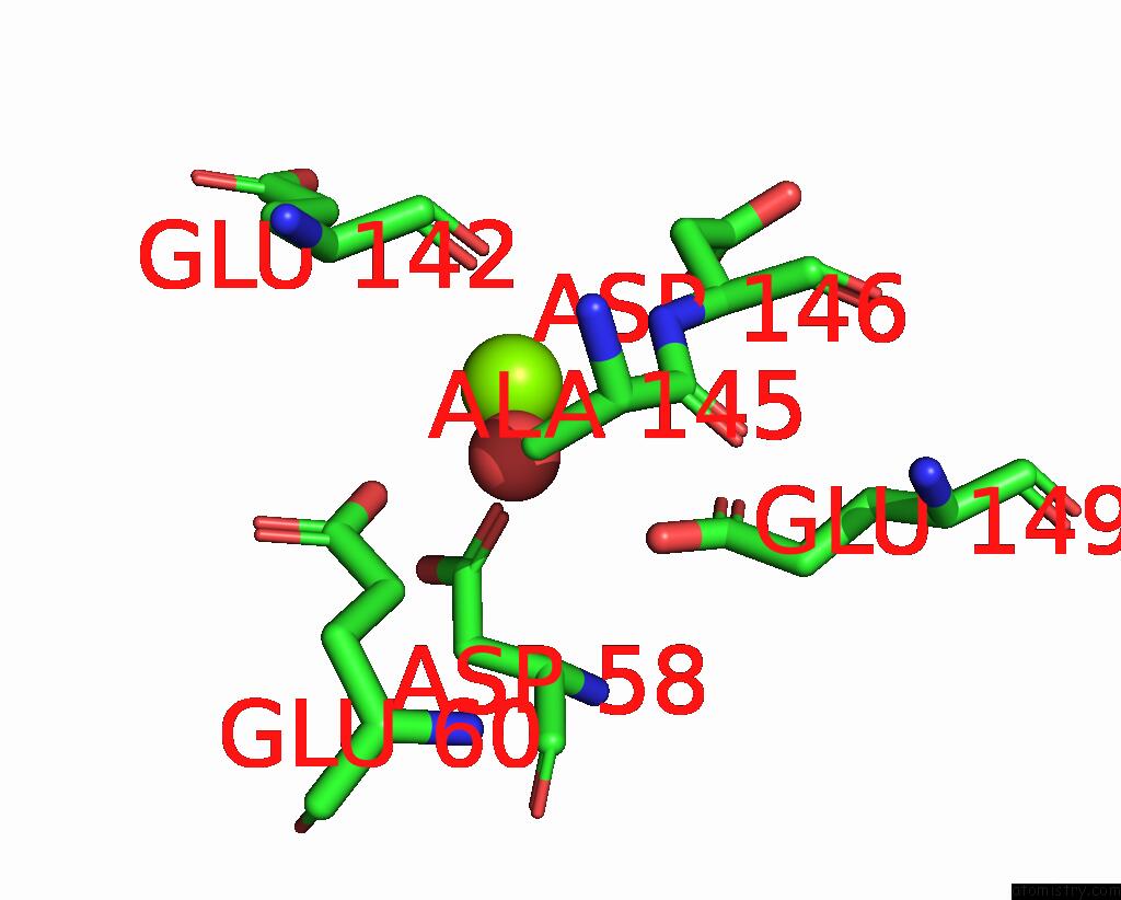
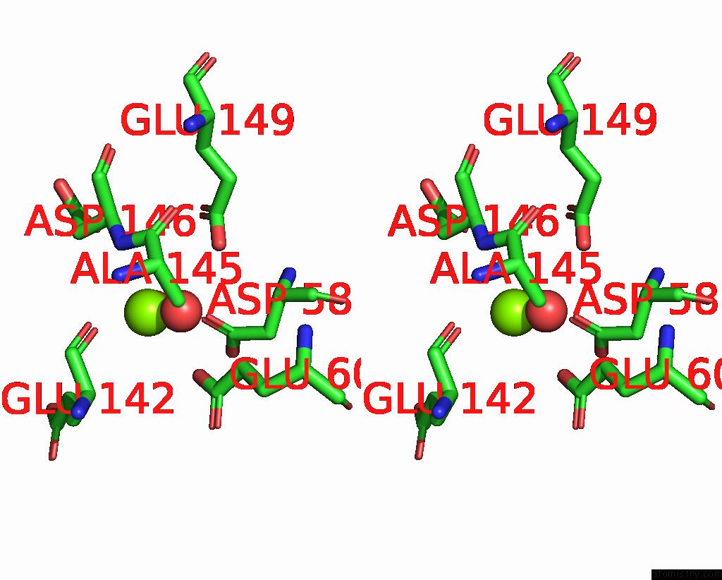
Magnesium binding site 1 out of 7 in 5n77
Go back to
Magnesium binding site 1 out
of 7 in the Crystal Structure of the Cytosolic Domain of the Cora Magnesium Channel From Escherichia Coli in Complex with Magnesium
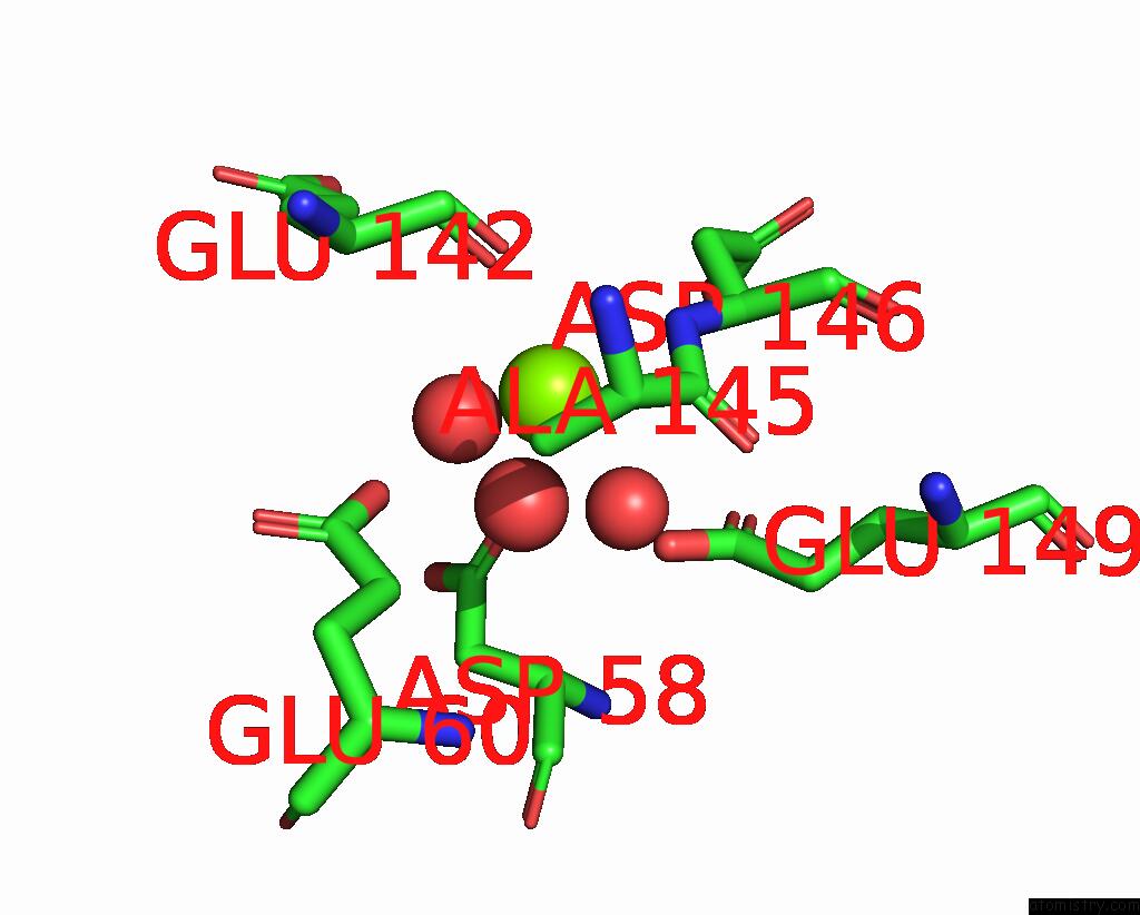
Mono view
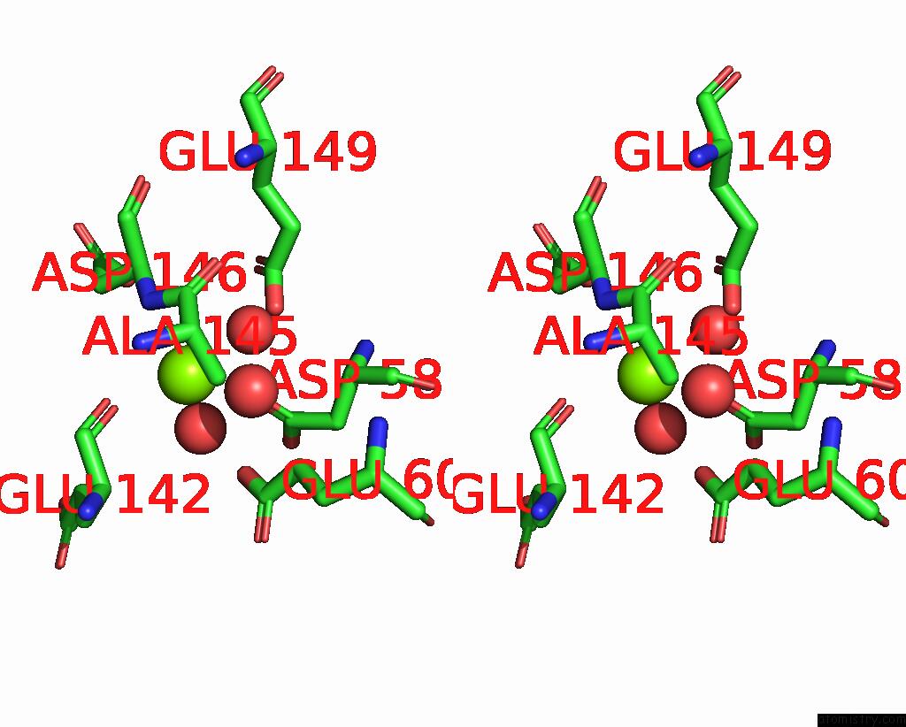
Stereo pair view
Mono view
Stereo pair view
A full contact list of Magnesium with other atoms in the Mg binding
site number 1 of Crystal Structure of the Cytosolic Domain of the Cora Magnesium Channel From Escherichia Coli in Complex with Magnesium within 5.0Å range:
|
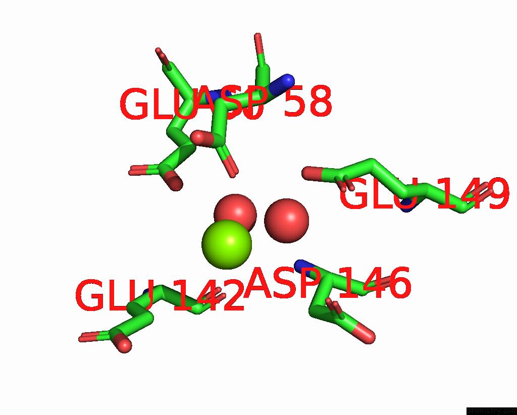
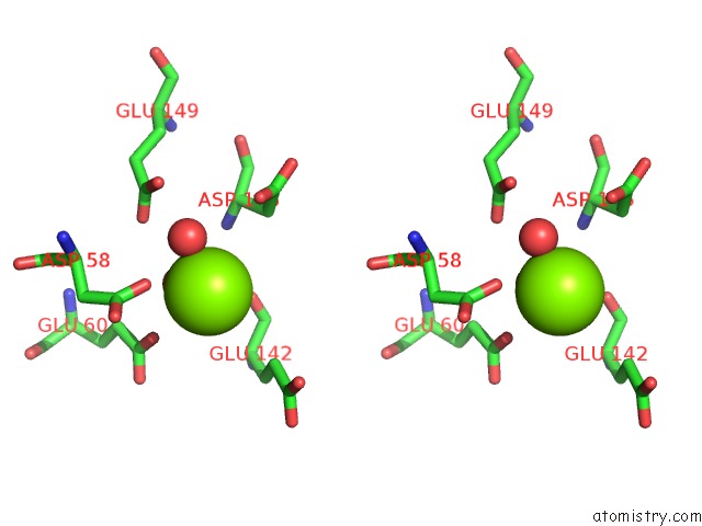
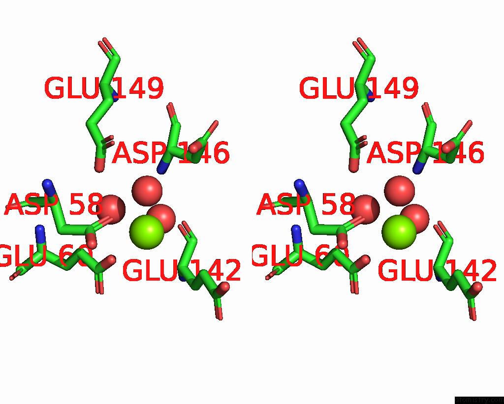
Magnesium binding site 2 out of 7 in 5n77
Go back to
Magnesium binding site 2 out
of 7 in the Crystal Structure of the Cytosolic Domain of the Cora Magnesium Channel From Escherichia Coli in Complex with Magnesium
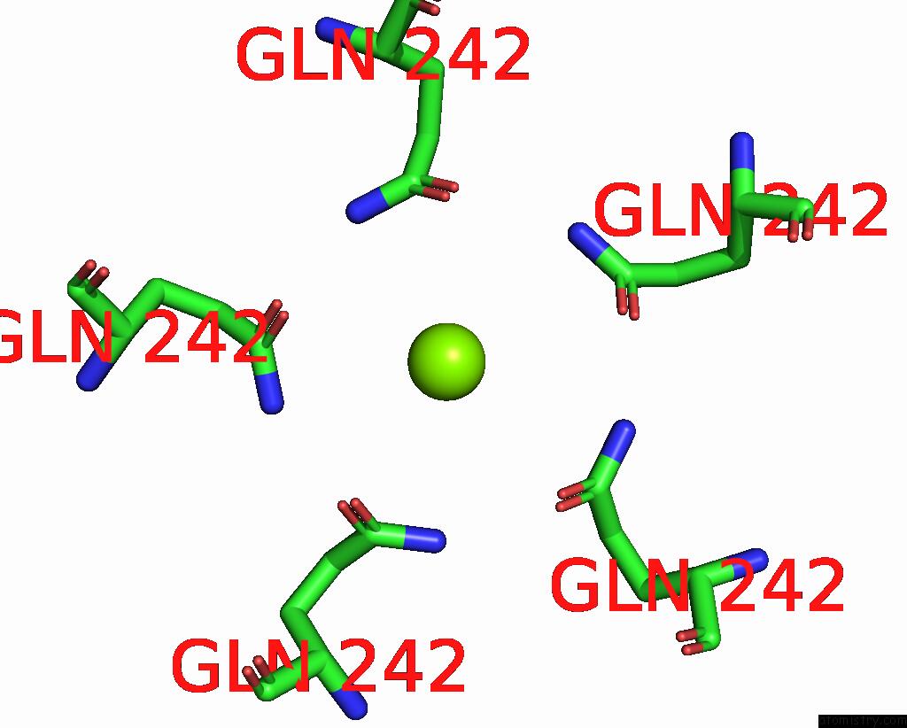
Mono view
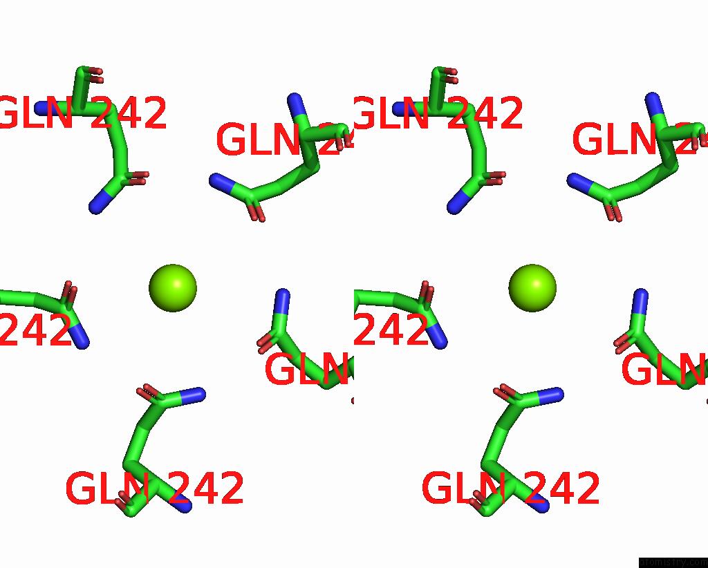
Stereo pair view
Mono view
Stereo pair view
A full contact list of Magnesium with other atoms in the Mg binding
site number 2 of Crystal Structure of the Cytosolic Domain of the Cora Magnesium Channel From Escherichia Coli in Complex with Magnesium within 5.0Å range:
|
Magnesium binding site 3 out of 7 in 5n77
Go back to
Magnesium binding site 3 out
of 7 in the Crystal Structure of the Cytosolic Domain of the Cora Magnesium Channel From Escherichia Coli in Complex with Magnesium
Mono view
Stereo pair view
Mono view
Stereo pair view
A full contact list of Magnesium with other atoms in the Mg binding
site number 3 of Crystal Structure of the Cytosolic Domain of the Cora Magnesium Channel From Escherichia Coli in Complex with Magnesium within 5.0Å range:
|
Magnesium binding site 4 out of 7 in 5n77
Go back to
Magnesium binding site 4 out
of 7 in the Crystal Structure of the Cytosolic Domain of the Cora Magnesium Channel From Escherichia Coli in Complex with Magnesium
Mono view
Stereo pair view
Mono view
Stereo pair view
A full contact list of Magnesium with other atoms in the Mg binding
site number 4 of Crystal Structure of the Cytosolic Domain of the Cora Magnesium Channel From Escherichia Coli in Complex with Magnesium within 5.0Å range:
|
Magnesium binding site 5 out of 7 in 5n77
Go back to
Magnesium binding site 5 out
of 7 in the Crystal Structure of the Cytosolic Domain of the Cora Magnesium Channel From Escherichia Coli in Complex with Magnesium
Mono view
Stereo pair view
Mono view
Stereo pair view
A full contact list of Magnesium with other atoms in the Mg binding
site number 5 of Crystal Structure of the Cytosolic Domain of the Cora Magnesium Channel From Escherichia Coli in Complex with Magnesium within 5.0Å range:
|
Magnesium binding site 6 out of 7 in 5n77
Go back to
Magnesium binding site 6 out
of 7 in the Crystal Structure of the Cytosolic Domain of the Cora Magnesium Channel From Escherichia Coli in Complex with Magnesium

Mono view

Stereo pair view
Mono view
Stereo pair view
A full contact list of Magnesium with other atoms in the Mg binding
site number 6 of Crystal Structure of the Cytosolic Domain of the Cora Magnesium Channel From Escherichia Coli in Complex with Magnesium within 5.0Å range:
|
Magnesium binding site 7 out of 7 in 5n77
Go back to
Magnesium binding site 7 out
of 7 in the Crystal Structure of the Cytosolic Domain of the Cora Magnesium Channel From Escherichia Coli in Complex with Magnesium

Mono view
Stereo pair view
Mono view
Stereo pair view
A full contact list of Magnesium with other atoms in the Mg binding
site number 7 of Crystal Structure of the Cytosolic Domain of the Cora Magnesium Channel From Escherichia Coli in Complex with Magnesium within 5.0Å range:
|
Reference:
M.Lerche,
H.Sandhu,
L.Flockner,
M.Hogbom,
M.Rapp.
Structure and Cooperativity of the Cytosolic Domain of the Cora Mg(2+) Channel From Escherichia Coli. Structure V. 25 1175 2017.
ISSN: ISSN 1878-4186
PubMed: 28669631
DOI: 10.1016/J.STR.2017.05.024
Page generated: Sun Sep 29 23:07:57 2024
ISSN: ISSN 1878-4186
PubMed: 28669631
DOI: 10.1016/J.STR.2017.05.024
Last articles
Zn in 9J0NZn in 9J0O
Zn in 9J0P
Zn in 9FJX
Zn in 9EKB
Zn in 9C0F
Zn in 9CAH
Zn in 9CH0
Zn in 9CH3
Zn in 9CH1