Magnesium »
PDB 5n96-5nol »
5nh7 »
Magnesium in PDB 5nh7: Crystal Structure of Xylose Isomerase From Piromyces E2 in Complex with Two MG2+ Ions and Xylose
Enzymatic activity of Crystal Structure of Xylose Isomerase From Piromyces E2 in Complex with Two MG2+ Ions and Xylose
All present enzymatic activity of Crystal Structure of Xylose Isomerase From Piromyces E2 in Complex with Two MG2+ Ions and Xylose:
5.3.1.5;
5.3.1.5;
Protein crystallography data
The structure of Crystal Structure of Xylose Isomerase From Piromyces E2 in Complex with Two MG2+ Ions and Xylose, PDB code: 5nh7
was solved by
H.J.Rozeboom,
D.B.Janssen,
with X-Ray Crystallography technique. A brief refinement statistics is given in the table below:
| Resolution Low / High (Å) | 39.50 / 1.90 |
| Space group | P 1 |
| Cell size a, b, c (Å), α, β, γ (°) | 78.600, 79.280, 91.969, 115.40, 89.91, 117.13 |
| R / Rfree (%) | 15.3 / 17.6 |
Magnesium Binding Sites:
Pages:
>>> Page 1 <<< Page 2, Binding sites: 11 - 12;Binding sites:
The binding sites of Magnesium atom in the Crystal Structure of Xylose Isomerase From Piromyces E2 in Complex with Two MG2+ Ions and Xylose (pdb code 5nh7). This binding sites where shown within 5.0 Angstroms radius around Magnesium atom.In total 12 binding sites of Magnesium where determined in the Crystal Structure of Xylose Isomerase From Piromyces E2 in Complex with Two MG2+ Ions and Xylose, PDB code: 5nh7:
Jump to Magnesium binding site number: 1; 2; 3; 4; 5; 6; 7; 8; 9; 10;
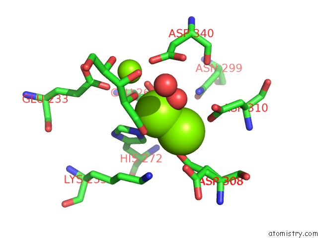
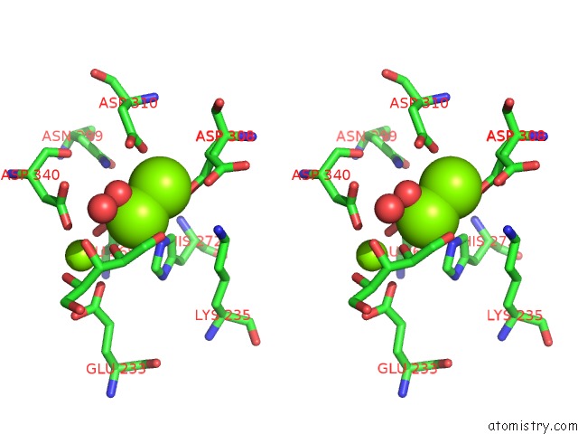
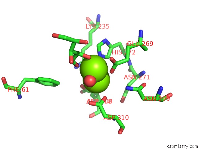
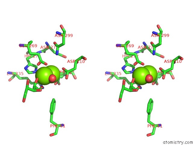
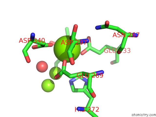
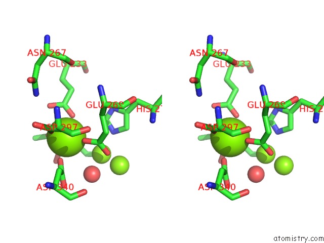
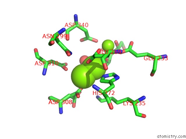
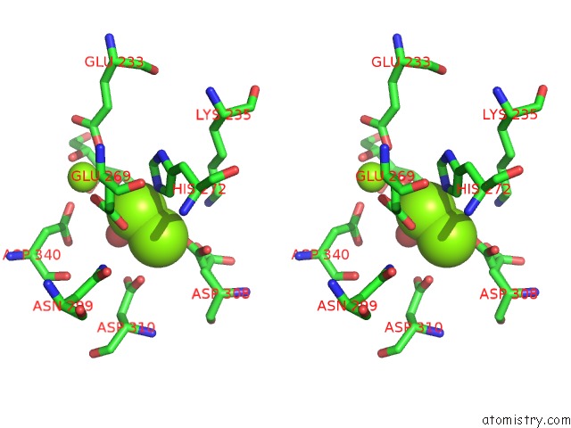
Magnesium binding site 1 out of 12 in 5nh7
Go back to
Magnesium binding site 1 out
of 12 in the Crystal Structure of Xylose Isomerase From Piromyces E2 in Complex with Two MG2+ Ions and Xylose
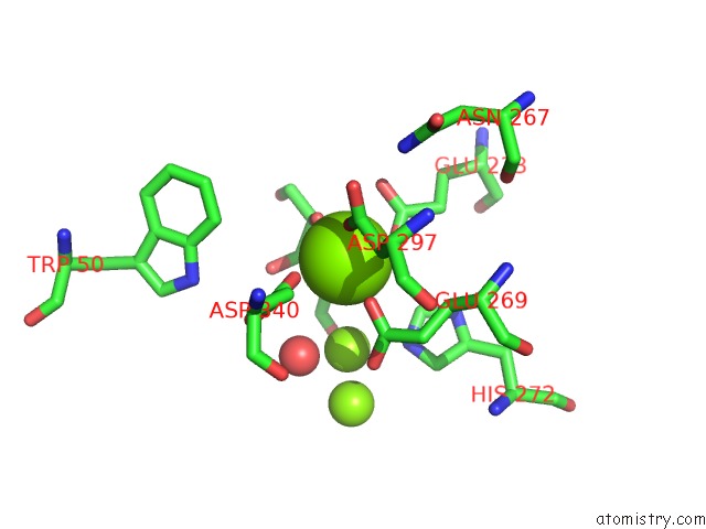
Mono view
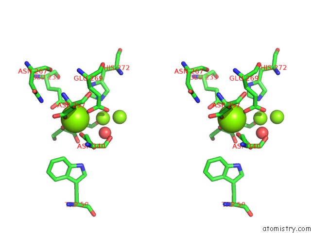
Stereo pair view
Mono view
Stereo pair view
A full contact list of Magnesium with other atoms in the Mg binding
site number 1 of Crystal Structure of Xylose Isomerase From Piromyces E2 in Complex with Two MG2+ Ions and Xylose within 5.0Å range:
|
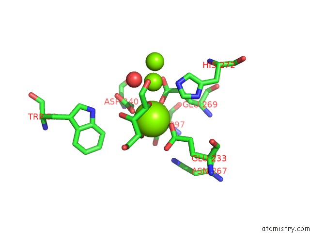
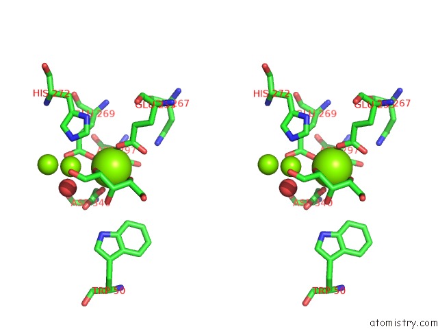
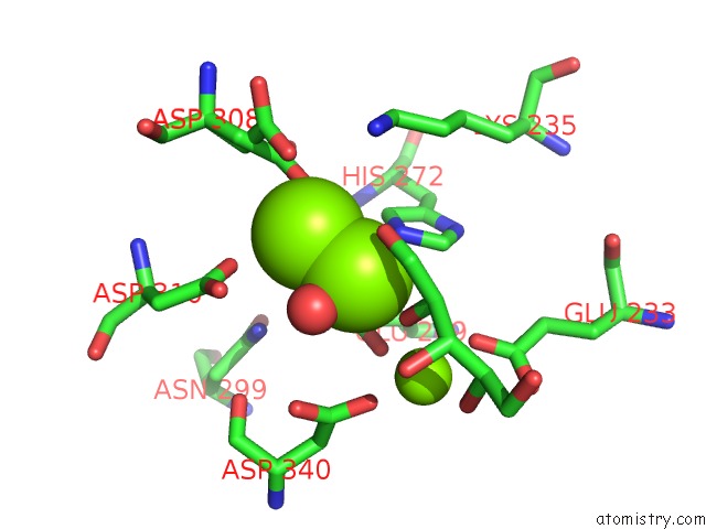
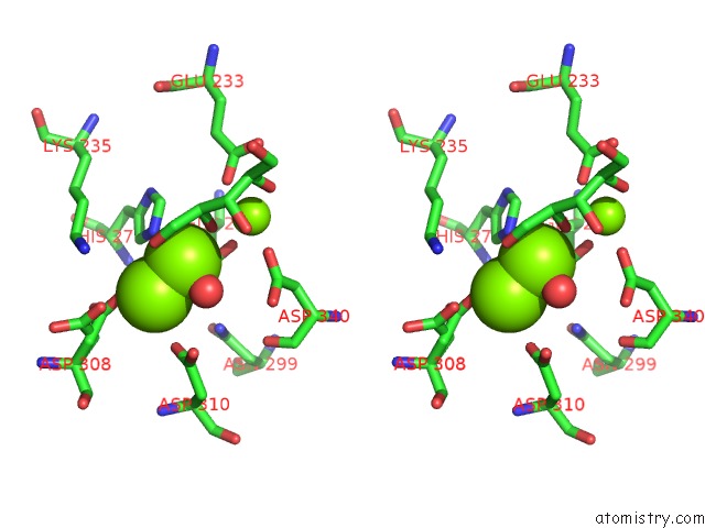
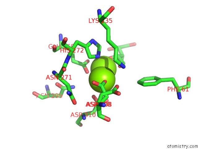
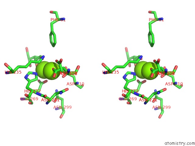
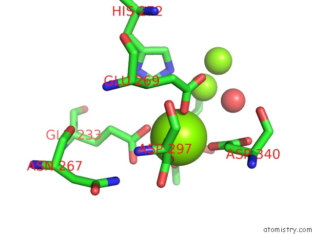
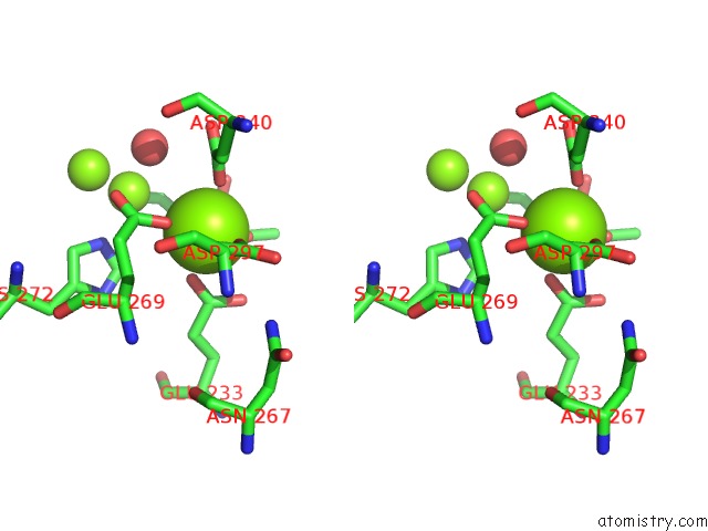
Magnesium binding site 2 out of 12 in 5nh7
Go back to
Magnesium binding site 2 out
of 12 in the Crystal Structure of Xylose Isomerase From Piromyces E2 in Complex with Two MG2+ Ions and Xylose
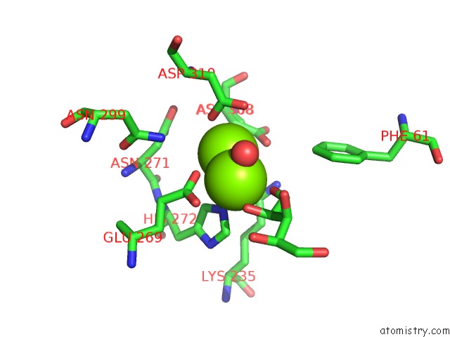
Mono view
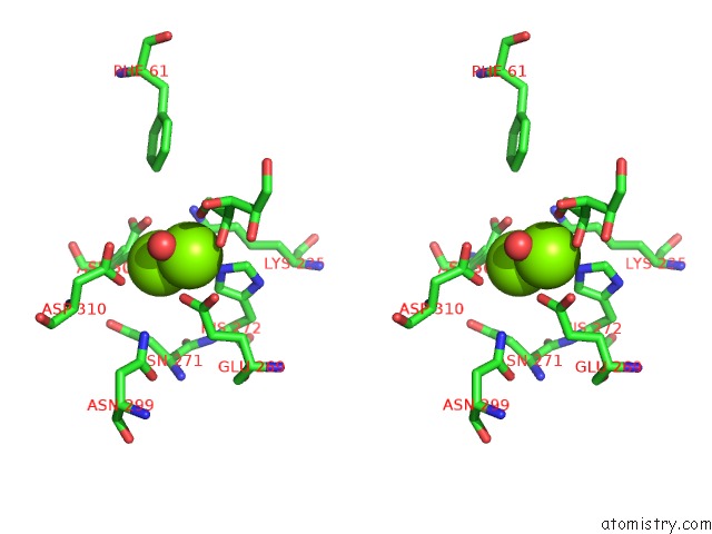
Stereo pair view
Mono view
Stereo pair view
A full contact list of Magnesium with other atoms in the Mg binding
site number 2 of Crystal Structure of Xylose Isomerase From Piromyces E2 in Complex with Two MG2+ Ions and Xylose within 5.0Å range:
|
Magnesium binding site 3 out of 12 in 5nh7
Go back to
Magnesium binding site 3 out
of 12 in the Crystal Structure of Xylose Isomerase From Piromyces E2 in Complex with Two MG2+ Ions and Xylose
Mono view
Stereo pair view
Mono view
Stereo pair view
A full contact list of Magnesium with other atoms in the Mg binding
site number 3 of Crystal Structure of Xylose Isomerase From Piromyces E2 in Complex with Two MG2+ Ions and Xylose within 5.0Å range:
|
Magnesium binding site 4 out of 12 in 5nh7
Go back to
Magnesium binding site 4 out
of 12 in the Crystal Structure of Xylose Isomerase From Piromyces E2 in Complex with Two MG2+ Ions and Xylose
Mono view
Stereo pair view
Mono view
Stereo pair view
A full contact list of Magnesium with other atoms in the Mg binding
site number 4 of Crystal Structure of Xylose Isomerase From Piromyces E2 in Complex with Two MG2+ Ions and Xylose within 5.0Å range:
|
Magnesium binding site 5 out of 12 in 5nh7
Go back to
Magnesium binding site 5 out
of 12 in the Crystal Structure of Xylose Isomerase From Piromyces E2 in Complex with Two MG2+ Ions and Xylose
Mono view
Stereo pair view
Mono view
Stereo pair view
A full contact list of Magnesium with other atoms in the Mg binding
site number 5 of Crystal Structure of Xylose Isomerase From Piromyces E2 in Complex with Two MG2+ Ions and Xylose within 5.0Å range:
|
Magnesium binding site 6 out of 12 in 5nh7
Go back to
Magnesium binding site 6 out
of 12 in the Crystal Structure of Xylose Isomerase From Piromyces E2 in Complex with Two MG2+ Ions and Xylose
Mono view
Stereo pair view
Mono view
Stereo pair view
A full contact list of Magnesium with other atoms in the Mg binding
site number 6 of Crystal Structure of Xylose Isomerase From Piromyces E2 in Complex with Two MG2+ Ions and Xylose within 5.0Å range:
|
Magnesium binding site 7 out of 12 in 5nh7
Go back to
Magnesium binding site 7 out
of 12 in the Crystal Structure of Xylose Isomerase From Piromyces E2 in Complex with Two MG2+ Ions and Xylose
Mono view
Stereo pair view
Mono view
Stereo pair view
A full contact list of Magnesium with other atoms in the Mg binding
site number 7 of Crystal Structure of Xylose Isomerase From Piromyces E2 in Complex with Two MG2+ Ions and Xylose within 5.0Å range:
|
Magnesium binding site 8 out of 12 in 5nh7
Go back to
Magnesium binding site 8 out
of 12 in the Crystal Structure of Xylose Isomerase From Piromyces E2 in Complex with Two MG2+ Ions and Xylose
Mono view
Stereo pair view
Mono view
Stereo pair view
A full contact list of Magnesium with other atoms in the Mg binding
site number 8 of Crystal Structure of Xylose Isomerase From Piromyces E2 in Complex with Two MG2+ Ions and Xylose within 5.0Å range:
|
Magnesium binding site 9 out of 12 in 5nh7
Go back to
Magnesium binding site 9 out
of 12 in the Crystal Structure of Xylose Isomerase From Piromyces E2 in Complex with Two MG2+ Ions and Xylose
Mono view
Stereo pair view
Mono view
Stereo pair view
A full contact list of Magnesium with other atoms in the Mg binding
site number 9 of Crystal Structure of Xylose Isomerase From Piromyces E2 in Complex with Two MG2+ Ions and Xylose within 5.0Å range:
|
Magnesium binding site 10 out of 12 in 5nh7
Go back to
Magnesium binding site 10 out
of 12 in the Crystal Structure of Xylose Isomerase From Piromyces E2 in Complex with Two MG2+ Ions and Xylose
Mono view
Stereo pair view
Mono view
Stereo pair view
A full contact list of Magnesium with other atoms in the Mg binding
site number 10 of Crystal Structure of Xylose Isomerase From Piromyces E2 in Complex with Two MG2+ Ions and Xylose within 5.0Å range:
|
Reference:
M.Lee,
H.J.Rozeboom,
P.P.De Waal,
R.M.De Jong,
H.M.Dudek,
D.B.Janssen.
Metal Dependence of the Xylose Isomerase From Piromyces Sp. E2 Explored By Activity Profiling and Protein Crystallography. Biochemistry V. 56 5991 2017.
ISSN: ISSN 1520-4995
PubMed: 29045784
DOI: 10.1021/ACS.BIOCHEM.7B00777
Page generated: Sun Sep 29 23:17:40 2024
ISSN: ISSN 1520-4995
PubMed: 29045784
DOI: 10.1021/ACS.BIOCHEM.7B00777
Last articles
Zn in 9MJ5Zn in 9HNW
Zn in 9G0L
Zn in 9FNE
Zn in 9DZN
Zn in 9E0I
Zn in 9D32
Zn in 9DAK
Zn in 8ZXC
Zn in 8ZUF