Magnesium »
PDB 5nol-5o5w »
5nz3 »
Magnesium in PDB 5nz3: The Structure of the Thermobifida Fusca Guanidine III Riboswitch with Methylguanidine
Protein crystallography data
The structure of The Structure of the Thermobifida Fusca Guanidine III Riboswitch with Methylguanidine, PDB code: 5nz3
was solved by
L.Huang,
J.Wang,
D.M.J.Lilley,
with X-Ray Crystallography technique. A brief refinement statistics is given in the table below:
| Resolution Low / High (Å) | 48.78 / 2.06 |
| Space group | P 31 2 1 |
| Cell size a, b, c (Å), α, β, γ (°) | 83.491, 83.491, 66.071, 90.00, 90.00, 120.00 |
| R / Rfree (%) | 21.9 / 27.4 |
Other elements in 5nz3:
The structure of The Structure of the Thermobifida Fusca Guanidine III Riboswitch with Methylguanidine also contains other interesting chemical elements:
| Bromine | (Br) | 4 atoms |
| Sodium | (Na) | 4 atoms |
Magnesium Binding Sites:
The binding sites of Magnesium atom in the The Structure of the Thermobifida Fusca Guanidine III Riboswitch with Methylguanidine
(pdb code 5nz3). This binding sites where shown within
5.0 Angstroms radius around Magnesium atom.
In total 5 binding sites of Magnesium where determined in the The Structure of the Thermobifida Fusca Guanidine III Riboswitch with Methylguanidine, PDB code: 5nz3:
Jump to Magnesium binding site number: 1; 2; 3; 4; 5;
In total 5 binding sites of Magnesium where determined in the The Structure of the Thermobifida Fusca Guanidine III Riboswitch with Methylguanidine, PDB code: 5nz3:
Jump to Magnesium binding site number: 1; 2; 3; 4; 5;
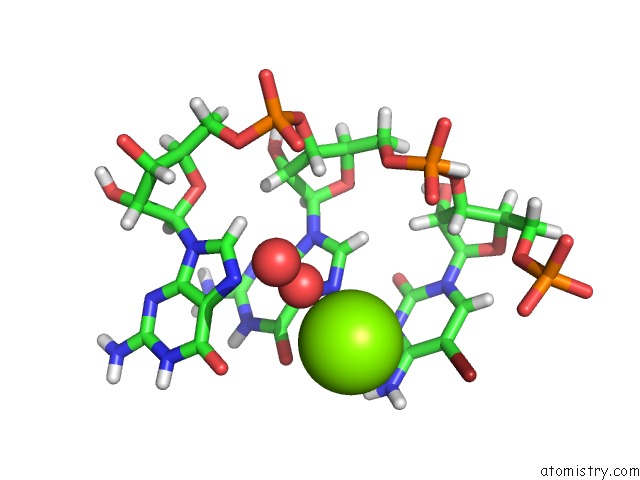
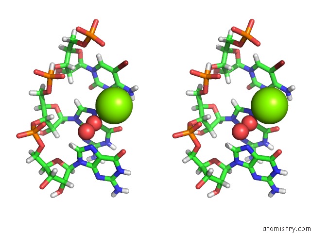
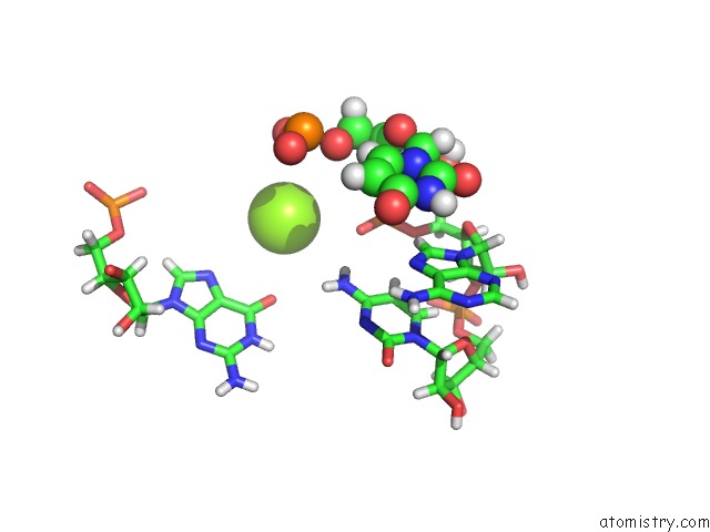
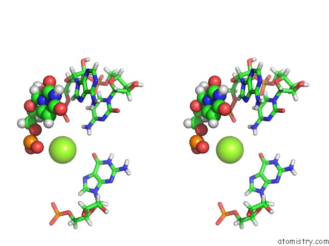
Magnesium binding site 1 out of 5 in 5nz3
Go back to
Magnesium binding site 1 out
of 5 in the The Structure of the Thermobifida Fusca Guanidine III Riboswitch with Methylguanidine
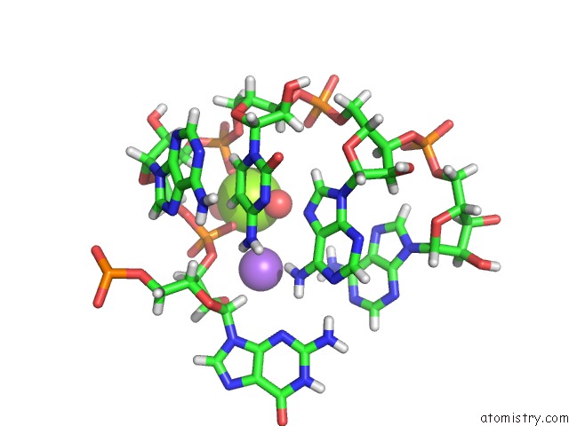
Mono view
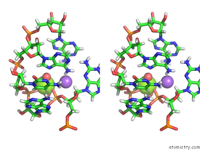
Stereo pair view
Mono view
Stereo pair view
A full contact list of Magnesium with other atoms in the Mg binding
site number 1 of The Structure of the Thermobifida Fusca Guanidine III Riboswitch with Methylguanidine within 5.0Å range:
|
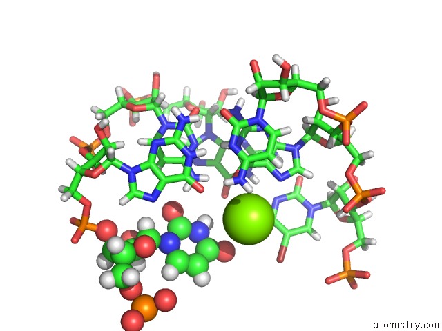
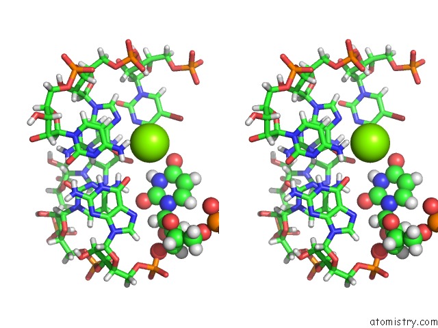
Magnesium binding site 2 out of 5 in 5nz3
Go back to
Magnesium binding site 2 out
of 5 in the The Structure of the Thermobifida Fusca Guanidine III Riboswitch with Methylguanidine
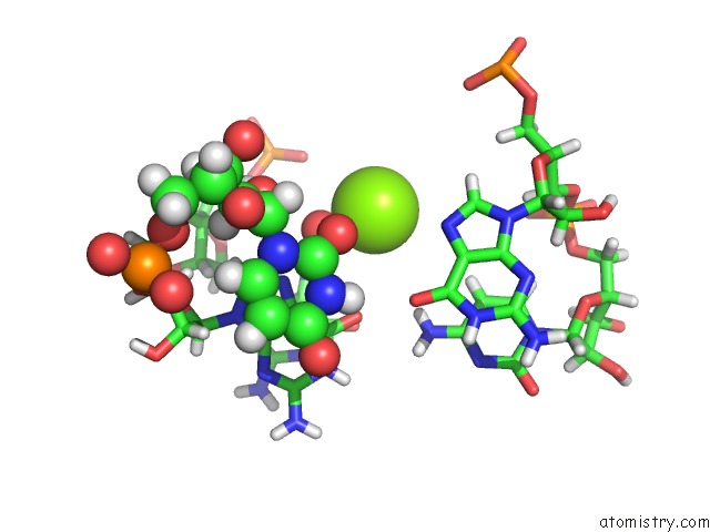
Mono view
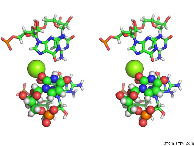
Stereo pair view
Mono view
Stereo pair view
A full contact list of Magnesium with other atoms in the Mg binding
site number 2 of The Structure of the Thermobifida Fusca Guanidine III Riboswitch with Methylguanidine within 5.0Å range:
|
Magnesium binding site 3 out of 5 in 5nz3
Go back to
Magnesium binding site 3 out
of 5 in the The Structure of the Thermobifida Fusca Guanidine III Riboswitch with Methylguanidine
Mono view
Stereo pair view
Mono view
Stereo pair view
A full contact list of Magnesium with other atoms in the Mg binding
site number 3 of The Structure of the Thermobifida Fusca Guanidine III Riboswitch with Methylguanidine within 5.0Å range:
|
Magnesium binding site 4 out of 5 in 5nz3
Go back to
Magnesium binding site 4 out
of 5 in the The Structure of the Thermobifida Fusca Guanidine III Riboswitch with Methylguanidine
Mono view
Stereo pair view
Mono view
Stereo pair view
A full contact list of Magnesium with other atoms in the Mg binding
site number 4 of The Structure of the Thermobifida Fusca Guanidine III Riboswitch with Methylguanidine within 5.0Å range:
|
Magnesium binding site 5 out of 5 in 5nz3
Go back to
Magnesium binding site 5 out
of 5 in the The Structure of the Thermobifida Fusca Guanidine III Riboswitch with Methylguanidine
Mono view
Stereo pair view
Mono view
Stereo pair view
A full contact list of Magnesium with other atoms in the Mg binding
site number 5 of The Structure of the Thermobifida Fusca Guanidine III Riboswitch with Methylguanidine within 5.0Å range:
|
Reference:
L.Huang,
J.Wang,
T.J.Wilson,
D.M.J.Lilley.
Structure of the Guanidine III Riboswitch. Cell Chem Biol V. 24 1407 2017.
ISSN: ESSN 2451-9456
PubMed: 28988949
DOI: 10.1016/J.CHEMBIOL.2017.08.021
Page generated: Sun Sep 29 23:43:26 2024
ISSN: ESSN 2451-9456
PubMed: 28988949
DOI: 10.1016/J.CHEMBIOL.2017.08.021
Last articles
Zn in 9J0NZn in 9J0O
Zn in 9J0P
Zn in 9FJX
Zn in 9EKB
Zn in 9C0F
Zn in 9CAH
Zn in 9CH0
Zn in 9CH3
Zn in 9CH1