Magnesium »
PDB 5o60-5odo »
5o7x »
Magnesium in PDB 5o7x: Crystal Structure of S. Cerevisiae Core Factor at 3.2A Resolution
Protein crystallography data
The structure of Crystal Structure of S. Cerevisiae Core Factor at 3.2A Resolution, PDB code: 5o7x
was solved by
C.Engel,
T.Gubbey,
S.Neyer,
S.Sainsbury,
C.Oberthuer,
C.Baejen,
C.Bernecky,
P.Cramer,
with X-Ray Crystallography technique. A brief refinement statistics is given in the table below:
| Resolution Low / High (Å) | 54.57 / 3.20 |
| Space group | P 1 |
| Cell size a, b, c (Å), α, β, γ (°) | 109.070, 109.140, 385.640, 90.02, 90.01, 59.98 |
| R / Rfree (%) | 25.4 / 28.3 |
Magnesium Binding Sites:
The binding sites of Magnesium atom in the Crystal Structure of S. Cerevisiae Core Factor at 3.2A Resolution
(pdb code 5o7x). This binding sites where shown within
5.0 Angstroms radius around Magnesium atom.
In total 6 binding sites of Magnesium where determined in the Crystal Structure of S. Cerevisiae Core Factor at 3.2A Resolution, PDB code: 5o7x:
Jump to Magnesium binding site number: 1; 2; 3; 4; 5; 6;
In total 6 binding sites of Magnesium where determined in the Crystal Structure of S. Cerevisiae Core Factor at 3.2A Resolution, PDB code: 5o7x:
Jump to Magnesium binding site number: 1; 2; 3; 4; 5; 6;
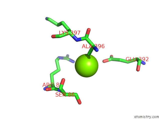
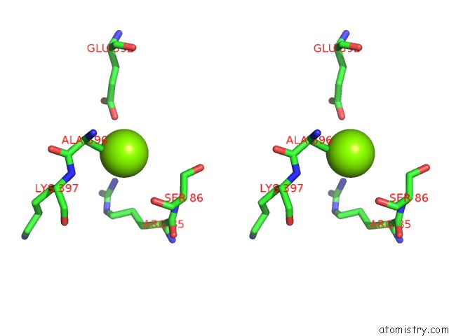
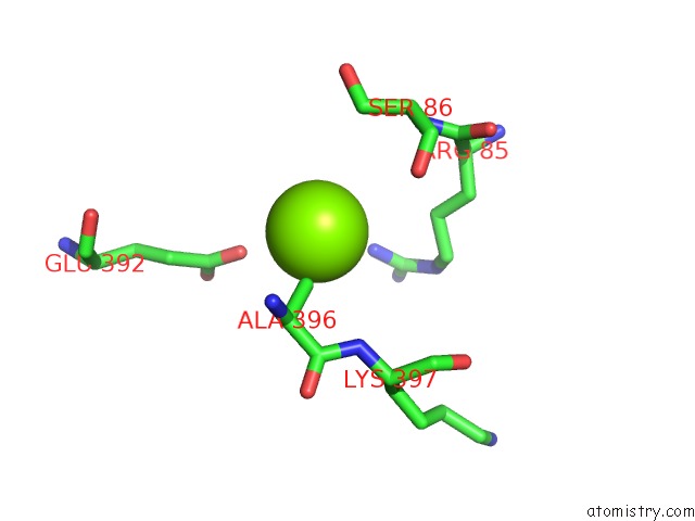
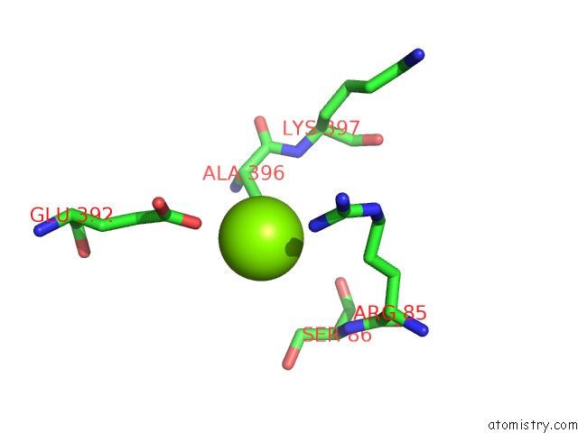
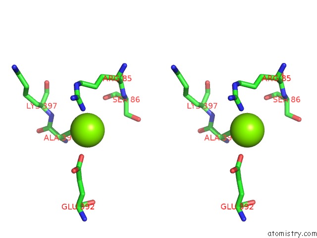
Magnesium binding site 1 out of 6 in 5o7x
Go back to
Magnesium binding site 1 out
of 6 in the Crystal Structure of S. Cerevisiae Core Factor at 3.2A Resolution
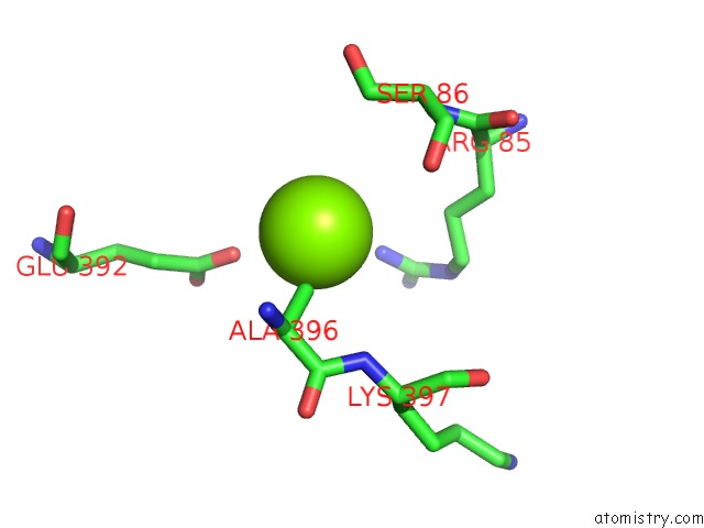
Mono view
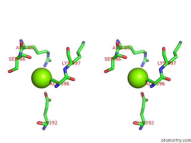
Stereo pair view
Mono view
Stereo pair view
A full contact list of Magnesium with other atoms in the Mg binding
site number 1 of Crystal Structure of S. Cerevisiae Core Factor at 3.2A Resolution within 5.0Å range:
|
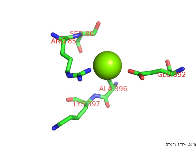
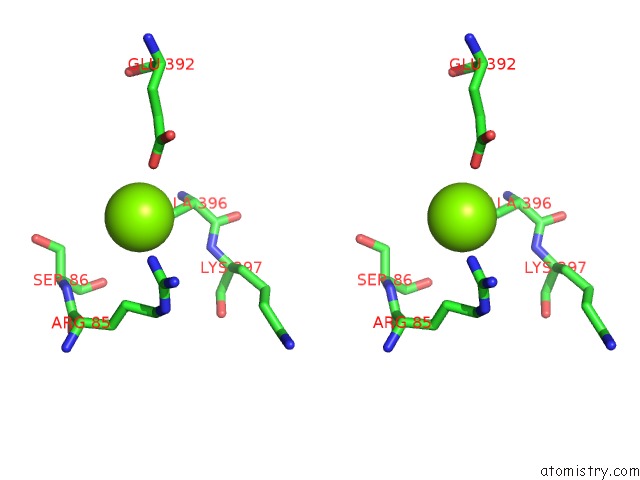
Magnesium binding site 2 out of 6 in 5o7x
Go back to
Magnesium binding site 2 out
of 6 in the Crystal Structure of S. Cerevisiae Core Factor at 3.2A Resolution
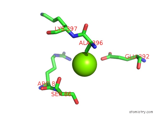
Mono view
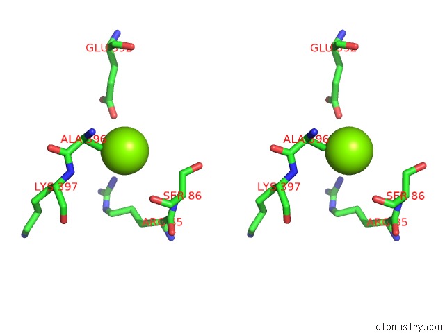
Stereo pair view
Mono view
Stereo pair view
A full contact list of Magnesium with other atoms in the Mg binding
site number 2 of Crystal Structure of S. Cerevisiae Core Factor at 3.2A Resolution within 5.0Å range:
|
Magnesium binding site 3 out of 6 in 5o7x
Go back to
Magnesium binding site 3 out
of 6 in the Crystal Structure of S. Cerevisiae Core Factor at 3.2A Resolution
Mono view
Stereo pair view
Mono view
Stereo pair view
A full contact list of Magnesium with other atoms in the Mg binding
site number 3 of Crystal Structure of S. Cerevisiae Core Factor at 3.2A Resolution within 5.0Å range:
|
Magnesium binding site 4 out of 6 in 5o7x
Go back to
Magnesium binding site 4 out
of 6 in the Crystal Structure of S. Cerevisiae Core Factor at 3.2A Resolution
Mono view
Stereo pair view
Mono view
Stereo pair view
A full contact list of Magnesium with other atoms in the Mg binding
site number 4 of Crystal Structure of S. Cerevisiae Core Factor at 3.2A Resolution within 5.0Å range:
|
Magnesium binding site 5 out of 6 in 5o7x
Go back to
Magnesium binding site 5 out
of 6 in the Crystal Structure of S. Cerevisiae Core Factor at 3.2A Resolution
Mono view

Stereo pair view
Mono view
Stereo pair view
A full contact list of Magnesium with other atoms in the Mg binding
site number 5 of Crystal Structure of S. Cerevisiae Core Factor at 3.2A Resolution within 5.0Å range:
|
Magnesium binding site 6 out of 6 in 5o7x
Go back to
Magnesium binding site 6 out
of 6 in the Crystal Structure of S. Cerevisiae Core Factor at 3.2A Resolution
Mono view
Stereo pair view
Mono view
Stereo pair view
A full contact list of Magnesium with other atoms in the Mg binding
site number 6 of Crystal Structure of S. Cerevisiae Core Factor at 3.2A Resolution within 5.0Å range:
|
Reference:
C.Engel,
T.Gubbey,
S.Neyer,
S.Sainsbury,
C.Oberthuer,
C.Baejen,
C.Bernecky,
P.Cramer.
Structural Basis of Rna Polymerase I Transcription Initiation. Cell V. 169 120 2017.
ISSN: ISSN 1097-4172
PubMed: 28340337
DOI: 10.1016/J.CELL.2017.03.003
Page generated: Mon Sep 30 00:13:29 2024
ISSN: ISSN 1097-4172
PubMed: 28340337
DOI: 10.1016/J.CELL.2017.03.003
Last articles
Zn in 9J0NZn in 9J0O
Zn in 9J0P
Zn in 9FJX
Zn in 9EKB
Zn in 9C0F
Zn in 9CAH
Zn in 9CH0
Zn in 9CH3
Zn in 9CH1