Magnesium »
PDB 5o62-5odz »
5oat »
Magnesium in PDB 5oat: PINK1 Structure
Protein crystallography data
The structure of PINK1 Structure, PDB code: 5oat
was solved by
A.Kumar,
J.Tamjar,
H.I.Woodroof,
O.G.Raimi,
A.Y.Waddell,
M.Peggie,
M.M.K.Muqit,
D.M.F.Van Aalten,
with X-Ray Crystallography technique. A brief refinement statistics is given in the table below:
| Resolution Low / High (Å) | 178.84 / 2.78 |
| Space group | P 1 21 1 |
| Cell size a, b, c (Å), α, β, γ (°) | 84.915, 116.736, 179.344, 90.00, 94.29, 90.00 |
| R / Rfree (%) | 20.5 / 24.6 |
Magnesium Binding Sites:
The binding sites of Magnesium atom in the PINK1 Structure
(pdb code 5oat). This binding sites where shown within
5.0 Angstroms radius around Magnesium atom.
In total 6 binding sites of Magnesium where determined in the PINK1 Structure, PDB code: 5oat:
Jump to Magnesium binding site number: 1; 2; 3; 4; 5; 6;
In total 6 binding sites of Magnesium where determined in the PINK1 Structure, PDB code: 5oat:
Jump to Magnesium binding site number: 1; 2; 3; 4; 5; 6;
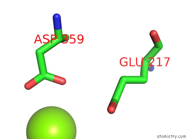
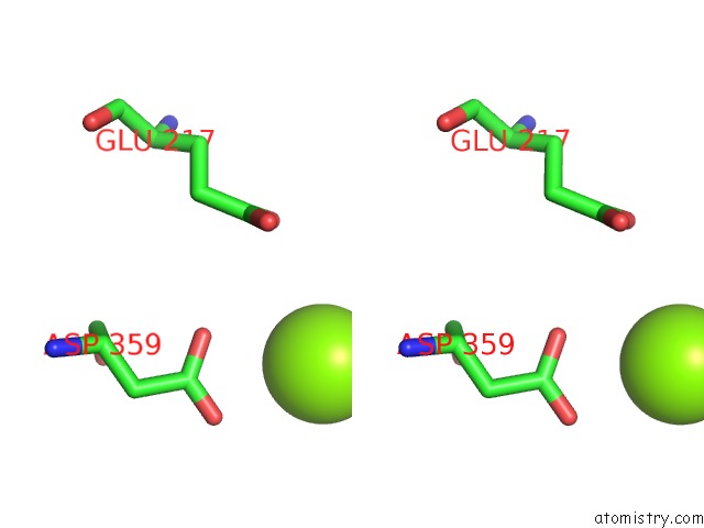
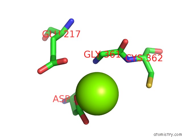
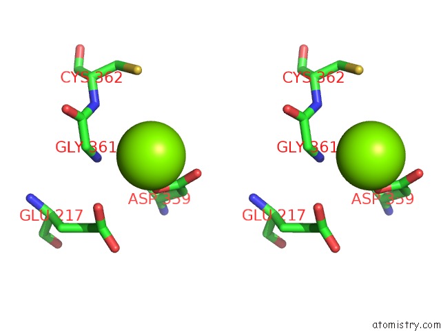
Magnesium binding site 1 out of 6 in 5oat
Go back to
Magnesium binding site 1 out
of 6 in the PINK1 Structure
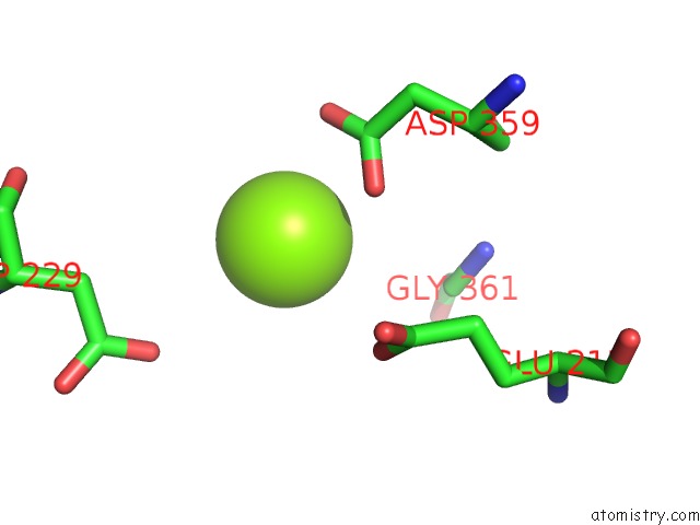
Mono view
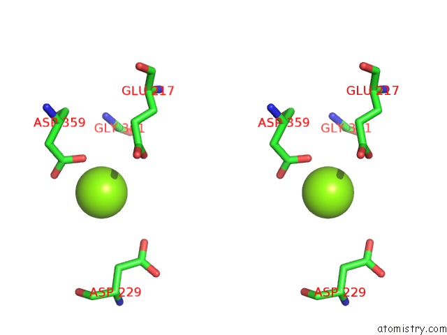
Stereo pair view
Mono view
Stereo pair view
A full contact list of Magnesium with other atoms in the Mg binding
site number 1 of PINK1 Structure within 5.0Å range:
|
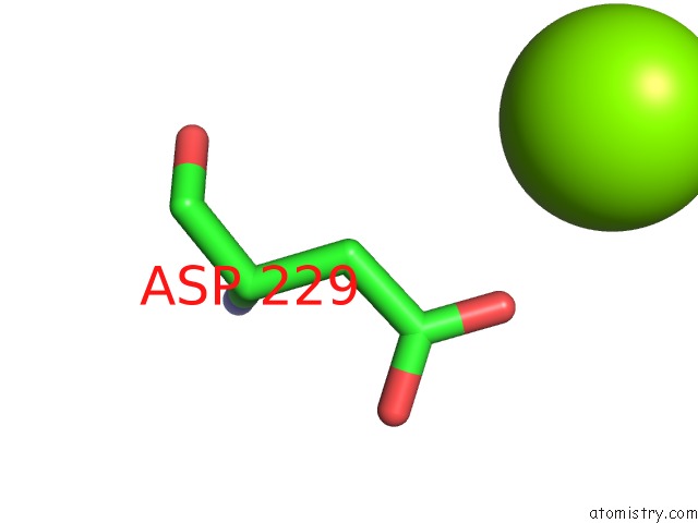
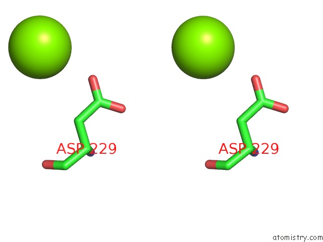
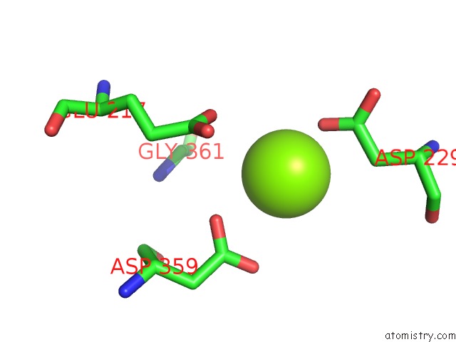
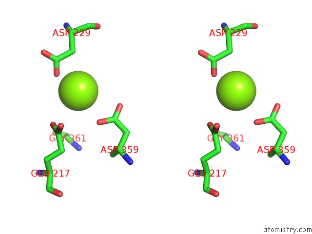
Magnesium binding site 2 out of 6 in 5oat
Go back to
Magnesium binding site 2 out
of 6 in the PINK1 Structure
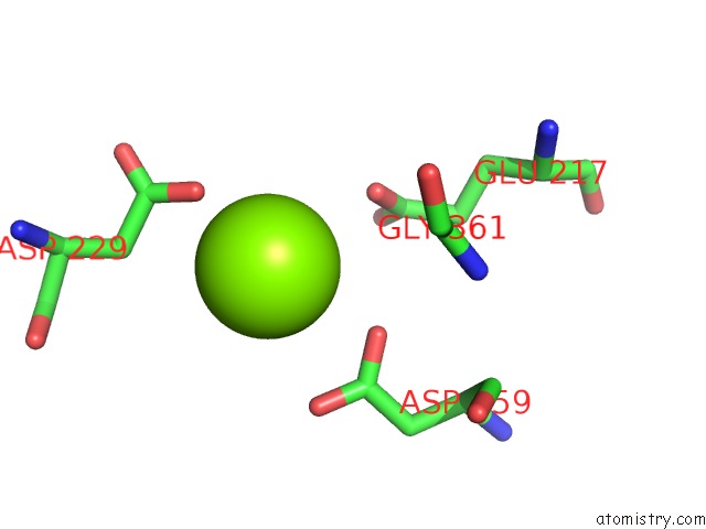
Mono view
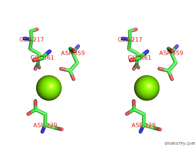
Stereo pair view
Mono view
Stereo pair view
A full contact list of Magnesium with other atoms in the Mg binding
site number 2 of PINK1 Structure within 5.0Å range:
|
Magnesium binding site 3 out of 6 in 5oat
Go back to
Magnesium binding site 3 out
of 6 in the PINK1 Structure
Mono view
Stereo pair view
Mono view
Stereo pair view
A full contact list of Magnesium with other atoms in the Mg binding
site number 3 of PINK1 Structure within 5.0Å range:
|
Magnesium binding site 4 out of 6 in 5oat
Go back to
Magnesium binding site 4 out
of 6 in the PINK1 Structure
Mono view
Stereo pair view
Mono view
Stereo pair view
A full contact list of Magnesium with other atoms in the Mg binding
site number 4 of PINK1 Structure within 5.0Å range:
|
Magnesium binding site 5 out of 6 in 5oat
Go back to
Magnesium binding site 5 out
of 6 in the PINK1 Structure
Mono view
Stereo pair view
Mono view
Stereo pair view
A full contact list of Magnesium with other atoms in the Mg binding
site number 5 of PINK1 Structure within 5.0Å range:
|
Magnesium binding site 6 out of 6 in 5oat
Go back to
Magnesium binding site 6 out
of 6 in the PINK1 Structure
Mono view
Stereo pair view
Mono view
Stereo pair view
A full contact list of Magnesium with other atoms in the Mg binding
site number 6 of PINK1 Structure within 5.0Å range:
|
Reference:
A.Kumar,
J.Tamjar,
A.D.Waddell,
H.I.Woodroof,
O.G.Raimi,
A.M.Shaw,
M.Peggie,
M.M.Muqit,
D.M.Van Aalten.
Structure of PINK1 and Mechanisms of Parkinson'S Disease Associated Mutations. Elife V. 6 2017.
ISSN: ESSN 2050-084X
PubMed: 28980524
DOI: 10.7554/ELIFE.29985
Page generated: Mon Sep 30 00:14:22 2024
ISSN: ESSN 2050-084X
PubMed: 28980524
DOI: 10.7554/ELIFE.29985
Last articles
Zn in 9MJ5Zn in 9HNW
Zn in 9G0L
Zn in 9FNE
Zn in 9DZN
Zn in 9E0I
Zn in 9D32
Zn in 9DAK
Zn in 8ZXC
Zn in 8ZUF