Magnesium »
PDB 5t3k-5tfc »
5t4y »
Magnesium in PDB 5t4y: Crystal Structure of BT1762-1763
Protein crystallography data
The structure of Crystal Structure of BT1762-1763, PDB code: 5t4y
was solved by
B.Van Den Berg,
with X-Ray Crystallography technique. A brief refinement statistics is given in the table below:
| Resolution Low / High (Å) | 130.35 / 3.10 |
| Space group | P 21 21 21 |
| Cell size a, b, c (Å), α, β, γ (°) | 110.826, 152.085, 253.005, 90.00, 90.00, 90.00 |
| R / Rfree (%) | 19.8 / 27 |
Magnesium Binding Sites:
The binding sites of Magnesium atom in the Crystal Structure of BT1762-1763
(pdb code 5t4y). This binding sites where shown within
5.0 Angstroms radius around Magnesium atom.
In total 5 binding sites of Magnesium where determined in the Crystal Structure of BT1762-1763, PDB code: 5t4y:
Jump to Magnesium binding site number: 1; 2; 3; 4; 5;
In total 5 binding sites of Magnesium where determined in the Crystal Structure of BT1762-1763, PDB code: 5t4y:
Jump to Magnesium binding site number: 1; 2; 3; 4; 5;
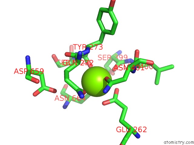
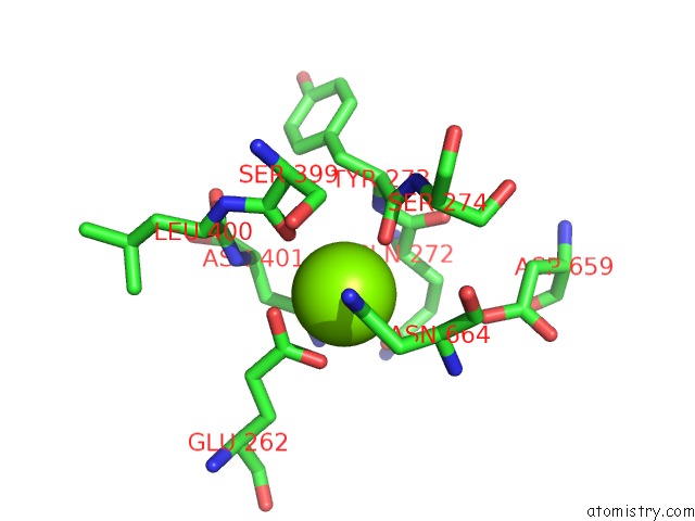
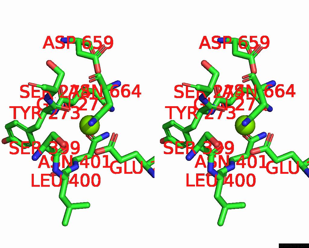
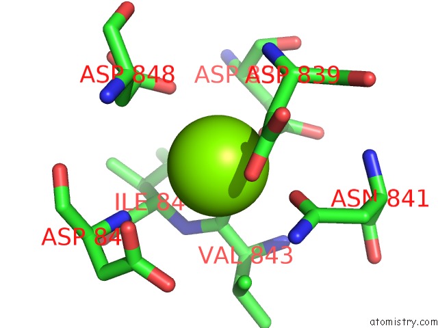
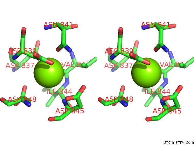
Magnesium binding site 1 out of 5 in 5t4y
Go back to
Magnesium binding site 1 out
of 5 in the Crystal Structure of BT1762-1763
Mono view
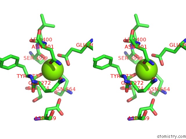
Stereo pair view
Mono view
Stereo pair view
A full contact list of Magnesium with other atoms in the Mg binding
site number 1 of Crystal Structure of BT1762-1763 within 5.0Å range:
|
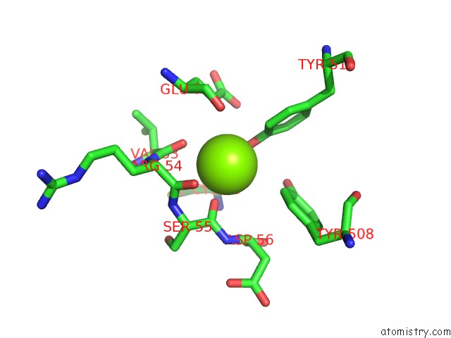
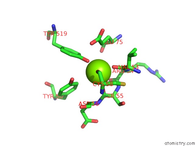
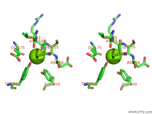
Magnesium binding site 2 out of 5 in 5t4y
Go back to
Magnesium binding site 2 out
of 5 in the Crystal Structure of BT1762-1763
Mono view
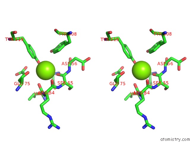
Stereo pair view
Mono view
Stereo pair view
A full contact list of Magnesium with other atoms in the Mg binding
site number 2 of Crystal Structure of BT1762-1763 within 5.0Å range:
|
Magnesium binding site 3 out of 5 in 5t4y
Go back to
Magnesium binding site 3 out
of 5 in the Crystal Structure of BT1762-1763
Mono view
Stereo pair view
Mono view
Stereo pair view
A full contact list of Magnesium with other atoms in the Mg binding
site number 3 of Crystal Structure of BT1762-1763 within 5.0Å range:
|
Magnesium binding site 4 out of 5 in 5t4y
Go back to
Magnesium binding site 4 out
of 5 in the Crystal Structure of BT1762-1763
Mono view
Stereo pair view
Mono view
Stereo pair view
A full contact list of Magnesium with other atoms in the Mg binding
site number 4 of Crystal Structure of BT1762-1763 within 5.0Å range:
|
Magnesium binding site 5 out of 5 in 5t4y
Go back to
Magnesium binding site 5 out
of 5 in the Crystal Structure of BT1762-1763
Mono view
Stereo pair view
Mono view
Stereo pair view
A full contact list of Magnesium with other atoms in the Mg binding
site number 5 of Crystal Structure of BT1762-1763 within 5.0Å range:
|
Reference:
A.J.Glenwright,
K.R.Pothula,
S.P.Bhamidimarri,
D.S.Chorev,
A.Basle,
S.J.Firbank,
H.Zheng,
C.V.Robinson,
M.Winterhalter,
U.Kleinekathofer,
D.N.Bolam,
B.Van Den Berg.
Structural Basis For Nutrient Acquisition By Dominant Members of the Human Gut Microbiota. Nature V. 541 407 2017.
ISSN: ESSN 1476-4687
PubMed: 28077872
DOI: 10.1038/NATURE20828
Page generated: Mon Sep 30 04:43:47 2024
ISSN: ESSN 1476-4687
PubMed: 28077872
DOI: 10.1038/NATURE20828
Last articles
F in 4GCAF in 4GE2
F in 4G9C
F in 4GAB
F in 4GA8
F in 4G90
F in 4G8R
F in 4G7G
F in 4G8O
F in 4G6O