Magnesium »
PDB 5t3k-5tfc »
5t9y »
Magnesium in PDB 5t9y: Crystal Structure of the Infectious Salmon Anemia Virus (Isav) Hemagglutinin-Esterase Protein
Protein crystallography data
The structure of Crystal Structure of the Infectious Salmon Anemia Virus (Isav) Hemagglutinin-Esterase Protein, PDB code: 5t9y
was solved by
J.D.Cook,
A.Sultana,
J.E.Lee,
with X-Ray Crystallography technique. A brief refinement statistics is given in the table below:
| Resolution Low / High (Å) | 47.72 / 1.80 |
| Space group | C 1 2 1 |
| Cell size a, b, c (Å), α, β, γ (°) | 142.419, 93.289, 96.722, 90.00, 122.01, 90.00 |
| R / Rfree (%) | 13.7 / 16.8 |
Magnesium Binding Sites:
The binding sites of Magnesium atom in the Crystal Structure of the Infectious Salmon Anemia Virus (Isav) Hemagglutinin-Esterase Protein
(pdb code 5t9y). This binding sites where shown within
5.0 Angstroms radius around Magnesium atom.
In total 3 binding sites of Magnesium where determined in the Crystal Structure of the Infectious Salmon Anemia Virus (Isav) Hemagglutinin-Esterase Protein, PDB code: 5t9y:
Jump to Magnesium binding site number: 1; 2; 3;
In total 3 binding sites of Magnesium where determined in the Crystal Structure of the Infectious Salmon Anemia Virus (Isav) Hemagglutinin-Esterase Protein, PDB code: 5t9y:
Jump to Magnesium binding site number: 1; 2; 3;
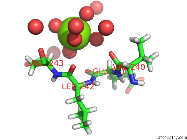
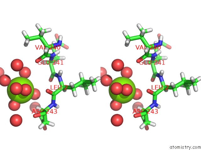
Magnesium binding site 1 out of 3 in 5t9y
Go back to
Magnesium binding site 1 out
of 3 in the Crystal Structure of the Infectious Salmon Anemia Virus (Isav) Hemagglutinin-Esterase Protein
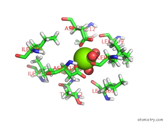
Mono view
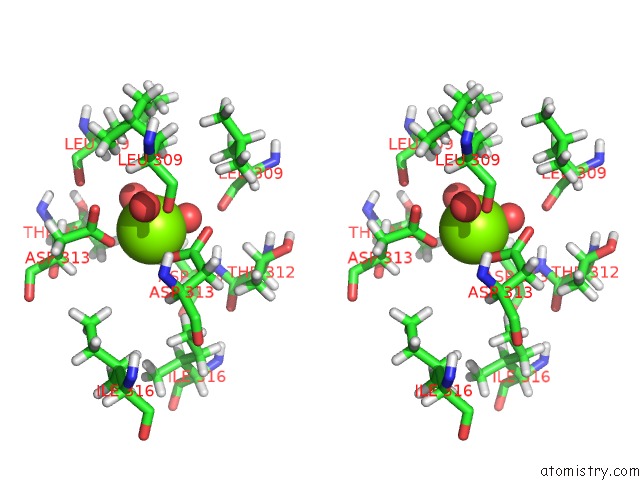
Stereo pair view
Mono view
Stereo pair view
A full contact list of Magnesium with other atoms in the Mg binding
site number 1 of Crystal Structure of the Infectious Salmon Anemia Virus (Isav) Hemagglutinin-Esterase Protein within 5.0Å range:
|
Magnesium binding site 2 out of 3 in 5t9y
Go back to
Magnesium binding site 2 out
of 3 in the Crystal Structure of the Infectious Salmon Anemia Virus (Isav) Hemagglutinin-Esterase Protein
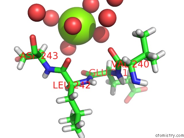
Mono view
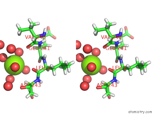
Stereo pair view
Mono view
Stereo pair view
A full contact list of Magnesium with other atoms in the Mg binding
site number 2 of Crystal Structure of the Infectious Salmon Anemia Virus (Isav) Hemagglutinin-Esterase Protein within 5.0Å range:
|
Magnesium binding site 3 out of 3 in 5t9y
Go back to
Magnesium binding site 3 out
of 3 in the Crystal Structure of the Infectious Salmon Anemia Virus (Isav) Hemagglutinin-Esterase Protein
Mono view
Stereo pair view
Mono view
Stereo pair view
A full contact list of Magnesium with other atoms in the Mg binding
site number 3 of Crystal Structure of the Infectious Salmon Anemia Virus (Isav) Hemagglutinin-Esterase Protein within 5.0Å range:
|
Reference:
J.D.Cook,
A.Sultana,
J.E.Lee.
Structure of the Infectious Salmon Anemia Virus Receptor Complex Illustrates A Unique Binding Strategy For Attachment. Proc. Natl. Acad. Sci. V. 114 E2929 2017U.S.A..
ISSN: ESSN 1091-6490
PubMed: 28320973
DOI: 10.1073/PNAS.1617993114
Page generated: Mon Sep 30 04:47:08 2024
ISSN: ESSN 1091-6490
PubMed: 28320973
DOI: 10.1073/PNAS.1617993114
Last articles
Fe in 2YXOFe in 2YRS
Fe in 2YXC
Fe in 2YNM
Fe in 2YVJ
Fe in 2YP1
Fe in 2YU2
Fe in 2YU1
Fe in 2YQB
Fe in 2YOO