Magnesium »
PDB 5tfd-5tus »
5thx »
Magnesium in PDB 5thx: Crystal Structure of A Ferredoxin Nadp+ Reductase From Neisseria Gonorrhoeae with Bound Nadp and Fad
Protein crystallography data
The structure of Crystal Structure of A Ferredoxin Nadp+ Reductase From Neisseria Gonorrhoeae with Bound Nadp and Fad, PDB code: 5thx
was solved by
Seattle Structural Genomics Center For Infectious Disease (Ssgcid),
with X-Ray Crystallography technique. A brief refinement statistics is given in the table below:
| Resolution Low / High (Å) | 37.40 / 1.55 |
| Space group | P 43 21 2 |
| Cell size a, b, c (Å), α, β, γ (°) | 60.010, 60.010, 158.270, 90.00, 90.00, 90.00 |
| R / Rfree (%) | 15.9 / 18.8 |
Magnesium Binding Sites:
The binding sites of Magnesium atom in the Crystal Structure of A Ferredoxin Nadp+ Reductase From Neisseria Gonorrhoeae with Bound Nadp and Fad
(pdb code 5thx). This binding sites where shown within
5.0 Angstroms radius around Magnesium atom.
In total only one binding site of Magnesium was determined in the Crystal Structure of A Ferredoxin Nadp+ Reductase From Neisseria Gonorrhoeae with Bound Nadp and Fad, PDB code: 5thx:
In total only one binding site of Magnesium was determined in the Crystal Structure of A Ferredoxin Nadp+ Reductase From Neisseria Gonorrhoeae with Bound Nadp and Fad, PDB code: 5thx:
Magnesium binding site 1 out of 1 in 5thx
Go back to
Magnesium binding site 1 out
of 1 in the Crystal Structure of A Ferredoxin Nadp+ Reductase From Neisseria Gonorrhoeae with Bound Nadp and Fad
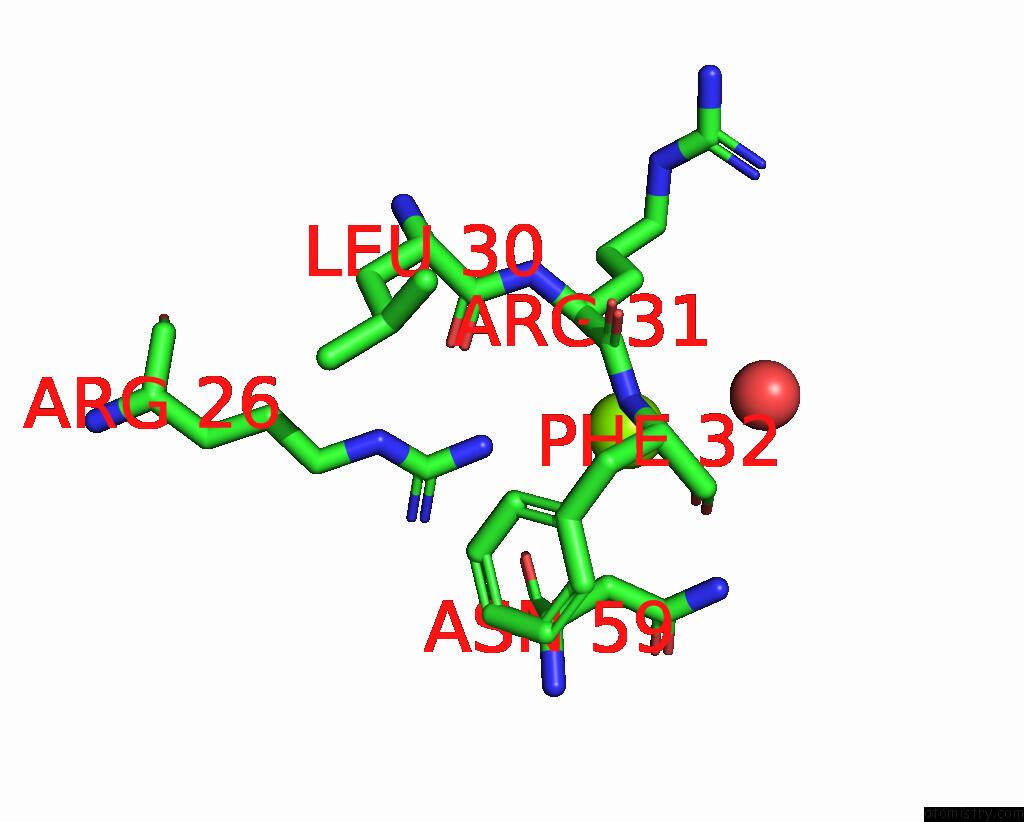
Mono view
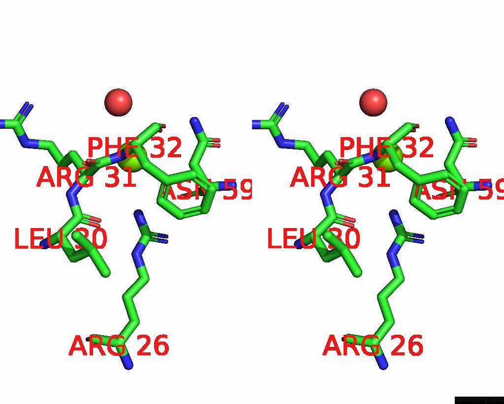
Stereo pair view
Mono view
Stereo pair view
A full contact list of Magnesium with other atoms in the Mg binding
site number 1 of Crystal Structure of A Ferredoxin Nadp+ Reductase From Neisseria Gonorrhoeae with Bound Nadp and Fad within 5.0Å range:
|
Reference:
J.N.Phan,
D.M.Dranow,
S.J.Mayclin,
D.D.Lorimer,
T.E.Edwards.
Crystal Structure of A Ferredoxin Nadp+ Reductase From Neisseria Gonorrhoeae with Bound Nadp and Fad To Be Published.
Page generated: Mon Sep 30 04:51:48 2024
Last articles
Cl in 5WIICl in 5WIU
Cl in 5WGZ
Cl in 5WGY
Cl in 5WHH
Cl in 5WGX
Cl in 5WGW
Cl in 5WGI
Cl in 5WGV
Cl in 5WGU