Magnesium »
PDB 5tfd-5tus »
5tk6 »
Magnesium in PDB 5tk6: Structure of the Hd-Domain Phosphohydrolase Oxsa with Oxetanocin-A Diphosphate Bound
Protein crystallography data
The structure of Structure of the Hd-Domain Phosphohydrolase Oxsa with Oxetanocin-A Diphosphate Bound, PDB code: 5tk6
was solved by
J.Bridwell-Rabb,
C.L.Drennan,
with X-Ray Crystallography technique. A brief refinement statistics is given in the table below:
| Resolution Low / High (Å) | 21.41 / 1.92 |
| Space group | H 3 2 |
| Cell size a, b, c (Å), α, β, γ (°) | 142.588, 142.588, 53.800, 90.00, 90.00, 120.00 |
| R / Rfree (%) | 17.9 / 21.8 |
Magnesium Binding Sites:
The binding sites of Magnesium atom in the Structure of the Hd-Domain Phosphohydrolase Oxsa with Oxetanocin-A Diphosphate Bound
(pdb code 5tk6). This binding sites where shown within
5.0 Angstroms radius around Magnesium atom.
In total 2 binding sites of Magnesium where determined in the Structure of the Hd-Domain Phosphohydrolase Oxsa with Oxetanocin-A Diphosphate Bound, PDB code: 5tk6:
Jump to Magnesium binding site number: 1; 2;
In total 2 binding sites of Magnesium where determined in the Structure of the Hd-Domain Phosphohydrolase Oxsa with Oxetanocin-A Diphosphate Bound, PDB code: 5tk6:
Jump to Magnesium binding site number: 1; 2;
Magnesium binding site 1 out of 2 in 5tk6
Go back to
Magnesium binding site 1 out
of 2 in the Structure of the Hd-Domain Phosphohydrolase Oxsa with Oxetanocin-A Diphosphate Bound
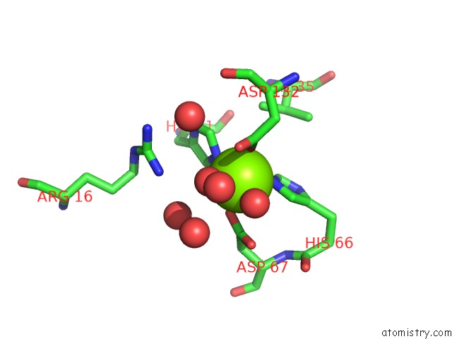
Mono view
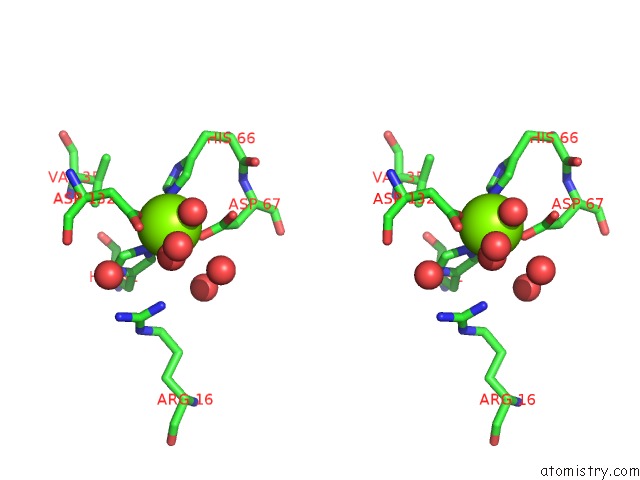
Stereo pair view
Mono view
Stereo pair view
A full contact list of Magnesium with other atoms in the Mg binding
site number 1 of Structure of the Hd-Domain Phosphohydrolase Oxsa with Oxetanocin-A Diphosphate Bound within 5.0Å range:
|
Magnesium binding site 2 out of 2 in 5tk6
Go back to
Magnesium binding site 2 out
of 2 in the Structure of the Hd-Domain Phosphohydrolase Oxsa with Oxetanocin-A Diphosphate Bound
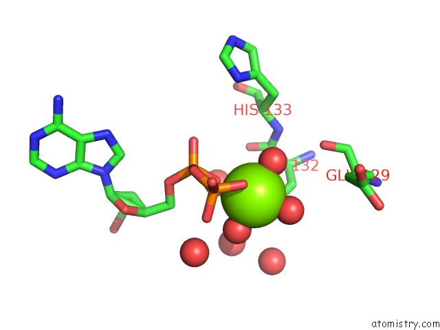
Mono view
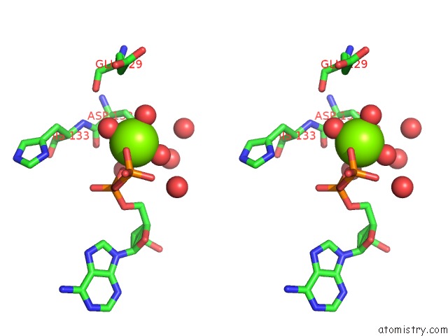
Stereo pair view
Mono view
Stereo pair view
A full contact list of Magnesium with other atoms in the Mg binding
site number 2 of Structure of the Hd-Domain Phosphohydrolase Oxsa with Oxetanocin-A Diphosphate Bound within 5.0Å range:
|
Reference:
J.Bridwell-Rabb,
G.Kang,
A.Zhong,
H.W.Liu,
C.L.Drennan.
An Hd Domain Phosphohydrolase Active Site Tailored For Oxetanocin-A Biosynthesis. Proc. Natl. Acad. Sci. V. 113 13750 2016U.S.A..
ISSN: ESSN 1091-6490
PubMed: 27849620
DOI: 10.1073/PNAS.1613610113
Page generated: Mon Sep 30 04:52:18 2024
ISSN: ESSN 1091-6490
PubMed: 27849620
DOI: 10.1073/PNAS.1613610113
Last articles
F in 4FF6F in 4FIM
F in 4FDO
F in 4FDN
F in 4FC0
F in 4FAT
F in 4F9Y
F in 4FA2
F in 4F9W
F in 4FAD