Magnesium »
PDB 5tfd-5tus »
5tk8 »
Magnesium in PDB 5tk8: Structure of the Hd-Domain Phosphohydrolase Oxsa with Oxetanocin-A Monophosphate Bound
Protein crystallography data
The structure of Structure of the Hd-Domain Phosphohydrolase Oxsa with Oxetanocin-A Monophosphate Bound, PDB code: 5tk8
was solved by
J.Bridwell-Rabb,
C.L.Drennan,
with X-Ray Crystallography technique. A brief refinement statistics is given in the table below:
| Resolution Low / High (Å) | 41.29 / 1.64 |
| Space group | H 3 2 |
| Cell size a, b, c (Å), α, β, γ (°) | 143.031, 143.031, 53.786, 90.00, 90.00, 120.00 |
| R / Rfree (%) | 17.9 / 20.4 |
Magnesium Binding Sites:
The binding sites of Magnesium atom in the Structure of the Hd-Domain Phosphohydrolase Oxsa with Oxetanocin-A Monophosphate Bound
(pdb code 5tk8). This binding sites where shown within
5.0 Angstroms radius around Magnesium atom.
In total only one binding site of Magnesium was determined in the Structure of the Hd-Domain Phosphohydrolase Oxsa with Oxetanocin-A Monophosphate Bound, PDB code: 5tk8:
In total only one binding site of Magnesium was determined in the Structure of the Hd-Domain Phosphohydrolase Oxsa with Oxetanocin-A Monophosphate Bound, PDB code: 5tk8:
Magnesium binding site 1 out of 1 in 5tk8
Go back to
Magnesium binding site 1 out
of 1 in the Structure of the Hd-Domain Phosphohydrolase Oxsa with Oxetanocin-A Monophosphate Bound
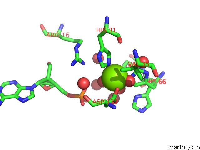
Mono view
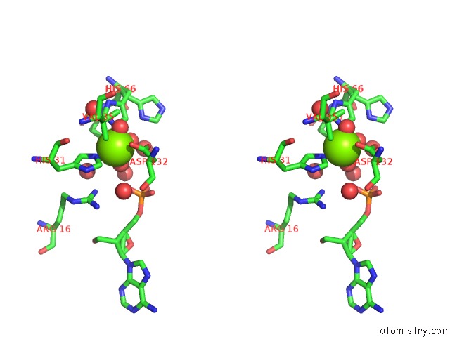
Stereo pair view
Mono view
Stereo pair view
A full contact list of Magnesium with other atoms in the Mg binding
site number 1 of Structure of the Hd-Domain Phosphohydrolase Oxsa with Oxetanocin-A Monophosphate Bound within 5.0Å range:
|
Reference:
J.Bridwell-Rabb,
G.Kang,
A.Zhong,
H.W.Liu,
C.L.Drennan.
An Hd Domain Phosphohydrolase Active Site Tailored For Oxetanocin-A Biosynthesis. Proc. Natl. Acad. Sci. V. 113 13750 2016U.S.A..
ISSN: ESSN 1091-6490
PubMed: 27849620
DOI: 10.1073/PNAS.1613610113
Page generated: Mon Sep 30 04:52:54 2024
ISSN: ESSN 1091-6490
PubMed: 27849620
DOI: 10.1073/PNAS.1613610113
Last articles
Cl in 5WL6Cl in 5WMD
Cl in 5WLY
Cl in 5WL3
Cl in 5WL7
Cl in 5WLG
Cl in 5WL5
Cl in 5WKX
Cl in 5WKY
Cl in 5WL1