Magnesium »
PDB 5u5q-5uhc »
5ual »
Magnesium in PDB 5ual: Escherichia Coli Rna Polymerase and Rifampin Complex, Rpob S531L Mutant
Enzymatic activity of Escherichia Coli Rna Polymerase and Rifampin Complex, Rpob S531L Mutant
All present enzymatic activity of Escherichia Coli Rna Polymerase and Rifampin Complex, Rpob S531L Mutant:
2.7.7.6;
2.7.7.6;
Protein crystallography data
The structure of Escherichia Coli Rna Polymerase and Rifampin Complex, Rpob S531L Mutant, PDB code: 5ual
was solved by
V.Molodtsov,
N.T.Scharf,
M.A.Stefan,
G.A.Garcia,
K.S.Murakami,
with X-Ray Crystallography technique. A brief refinement statistics is given in the table below:
| Resolution Low / High (Å) | 29.94 / 3.89 |
| Space group | P 21 21 21 |
| Cell size a, b, c (Å), α, β, γ (°) | 187.372, 205.950, 309.692, 90.00, 90.00, 90.00 |
| R / Rfree (%) | 23 / 28.6 |
Other elements in 5ual:
The structure of Escherichia Coli Rna Polymerase and Rifampin Complex, Rpob S531L Mutant also contains other interesting chemical elements:
| Zinc | (Zn) | 4 atoms |
Magnesium Binding Sites:
The binding sites of Magnesium atom in the Escherichia Coli Rna Polymerase and Rifampin Complex, Rpob S531L Mutant
(pdb code 5ual). This binding sites where shown within
5.0 Angstroms radius around Magnesium atom.
In total 2 binding sites of Magnesium where determined in the Escherichia Coli Rna Polymerase and Rifampin Complex, Rpob S531L Mutant, PDB code: 5ual:
Jump to Magnesium binding site number: 1; 2;
In total 2 binding sites of Magnesium where determined in the Escherichia Coli Rna Polymerase and Rifampin Complex, Rpob S531L Mutant, PDB code: 5ual:
Jump to Magnesium binding site number: 1; 2;
Magnesium binding site 1 out of 2 in 5ual
Go back to
Magnesium binding site 1 out
of 2 in the Escherichia Coli Rna Polymerase and Rifampin Complex, Rpob S531L Mutant
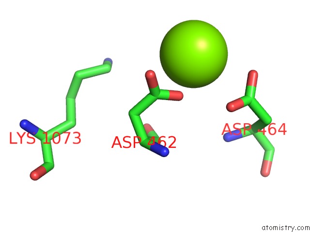
Mono view
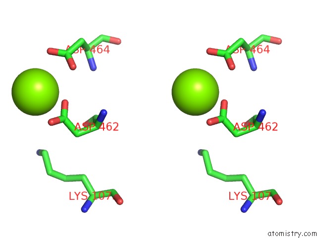
Stereo pair view
Mono view
Stereo pair view
A full contact list of Magnesium with other atoms in the Mg binding
site number 1 of Escherichia Coli Rna Polymerase and Rifampin Complex, Rpob S531L Mutant within 5.0Å range:
|
Magnesium binding site 2 out of 2 in 5ual
Go back to
Magnesium binding site 2 out
of 2 in the Escherichia Coli Rna Polymerase and Rifampin Complex, Rpob S531L Mutant
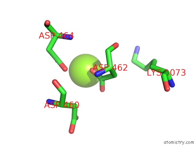
Mono view
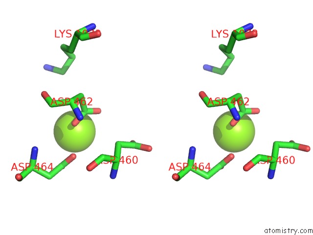
Stereo pair view
Mono view
Stereo pair view
A full contact list of Magnesium with other atoms in the Mg binding
site number 2 of Escherichia Coli Rna Polymerase and Rifampin Complex, Rpob S531L Mutant within 5.0Å range:
|
Reference:
V.Molodtsov,
N.T.Scharf,
M.A.Stefan,
G.A.Garcia,
K.S.Murakami.
Structural Basis For Rifamycin Resistance of Bacterial Rna Polymerase By the Three Most Clinically Important Rpob Mutations Found in Mycobacterium Tuberculosis. Mol. Microbiol. V. 103 1034 2017.
ISSN: ESSN 1365-2958
PubMed: 28009073
DOI: 10.1111/MMI.13606
Page generated: Mon Sep 30 05:15:45 2024
ISSN: ESSN 1365-2958
PubMed: 28009073
DOI: 10.1111/MMI.13606
Last articles
Fe in 2YXOFe in 2YRS
Fe in 2YXC
Fe in 2YNM
Fe in 2YVJ
Fe in 2YP1
Fe in 2YU2
Fe in 2YU1
Fe in 2YQB
Fe in 2YOO