Magnesium »
PDB 5usj-5v60 »
5uxc »
Magnesium in PDB 5uxc: Crystal Structure of Macrolide 2'-Phosphotransferase Mphh From Brachybacterium Faecium in Complex with Gdp
Protein crystallography data
The structure of Crystal Structure of Macrolide 2'-Phosphotransferase Mphh From Brachybacterium Faecium in Complex with Gdp, PDB code: 5uxc
was solved by
P.J.Stogios,
T.Skarina,
Z.Wawrzak,
V.Yim,
A.Savchenko,
W.F.Anderson,
Centerfor Structural Genomics Of Infectious Diseases (Csgid),
with X-Ray Crystallography technique. A brief refinement statistics is given in the table below:
| Resolution Low / High (Å) | 40.24 / 1.72 |
| Space group | P 21 21 21 |
| Cell size a, b, c (Å), α, β, γ (°) | 49.556, 77.002, 94.409, 90.00, 90.00, 90.00 |
| R / Rfree (%) | 16.4 / 20.7 |
Other elements in 5uxc:
The structure of Crystal Structure of Macrolide 2'-Phosphotransferase Mphh From Brachybacterium Faecium in Complex with Gdp also contains other interesting chemical elements:
| Chlorine | (Cl) | 2 atoms |
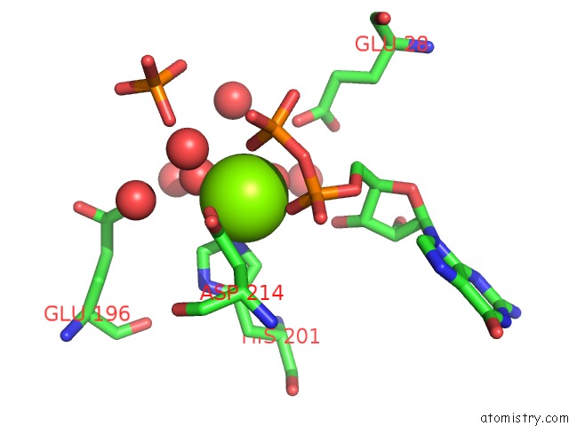
Magnesium Binding Sites:
The binding sites of Magnesium atom in the Crystal Structure of Macrolide 2'-Phosphotransferase Mphh From Brachybacterium Faecium in Complex with Gdp
(pdb code 5uxc). This binding sites where shown within
5.0 Angstroms radius around Magnesium atom.
In total only one binding site of Magnesium was determined in the Crystal Structure of Macrolide 2'-Phosphotransferase Mphh From Brachybacterium Faecium in Complex with Gdp, PDB code: 5uxc:
In total only one binding site of Magnesium was determined in the Crystal Structure of Macrolide 2'-Phosphotransferase Mphh From Brachybacterium Faecium in Complex with Gdp, PDB code: 5uxc:
Magnesium binding site 1 out of 1 in 5uxc
Go back to
Magnesium binding site 1 out
of 1 in the Crystal Structure of Macrolide 2'-Phosphotransferase Mphh From Brachybacterium Faecium in Complex with Gdp
Mono view

Stereo pair view
Mono view
Stereo pair view
A full contact list of Magnesium with other atoms in the Mg binding
site number 1 of Crystal Structure of Macrolide 2'-Phosphotransferase Mphh From Brachybacterium Faecium in Complex with Gdp within 5.0Å range:
|
Reference:
A.C.Pawlowski,
P.J.Stogios,
K.Koteva,
T.Skarina,
E.Evdokimova,
A.Savchenko,
G.D.Wright.
The Evolution of Substrate Discrimination in Macrolide Antibiotic Resistance Enzymes. Nat Commun V. 9 112 2018.
ISSN: ESSN 2041-1723
PubMed: 29317655
DOI: 10.1038/S41467-017-02680-0
Page generated: Mon Sep 30 05:27:13 2024
ISSN: ESSN 2041-1723
PubMed: 29317655
DOI: 10.1038/S41467-017-02680-0
Last articles
Zn in 9MJ5Zn in 9HNW
Zn in 9G0L
Zn in 9FNE
Zn in 9DZN
Zn in 9E0I
Zn in 9D32
Zn in 9DAK
Zn in 8ZXC
Zn in 8ZUF