Magnesium »
PDB 5v6s-5vp0 »
5vi8 »
Magnesium in PDB 5vi8: Structure of A Mycobacterium Smegmatis Transcription Initiation Complex with An Upstream-Fork Promoter Fragment
Enzymatic activity of Structure of A Mycobacterium Smegmatis Transcription Initiation Complex with An Upstream-Fork Promoter Fragment
All present enzymatic activity of Structure of A Mycobacterium Smegmatis Transcription Initiation Complex with An Upstream-Fork Promoter Fragment:
2.7.7.6;
2.7.7.6;
Protein crystallography data
The structure of Structure of A Mycobacterium Smegmatis Transcription Initiation Complex with An Upstream-Fork Promoter Fragment, PDB code: 5vi8
was solved by
E.A.Hubin,
E.A.Campbell,
S.A.Darst,
with X-Ray Crystallography technique. A brief refinement statistics is given in the table below:
| Resolution Low / High (Å) | 51.99 / 2.76 |
| Space group | P 1 21 1 |
| Cell size a, b, c (Å), α, β, γ (°) | 133.012, 161.633, 139.211, 90.00, 107.72, 90.00 |
| R / Rfree (%) | 24 / 28.1 |
Other elements in 5vi8:
The structure of Structure of A Mycobacterium Smegmatis Transcription Initiation Complex with An Upstream-Fork Promoter Fragment also contains other interesting chemical elements:
| Zinc | (Zn) | 2 atoms |
Magnesium Binding Sites:
The binding sites of Magnesium atom in the Structure of A Mycobacterium Smegmatis Transcription Initiation Complex with An Upstream-Fork Promoter Fragment
(pdb code 5vi8). This binding sites where shown within
5.0 Angstroms radius around Magnesium atom.
In total only one binding site of Magnesium was determined in the Structure of A Mycobacterium Smegmatis Transcription Initiation Complex with An Upstream-Fork Promoter Fragment, PDB code: 5vi8:
In total only one binding site of Magnesium was determined in the Structure of A Mycobacterium Smegmatis Transcription Initiation Complex with An Upstream-Fork Promoter Fragment, PDB code: 5vi8:
Magnesium binding site 1 out of 1 in 5vi8
Go back to
Magnesium binding site 1 out
of 1 in the Structure of A Mycobacterium Smegmatis Transcription Initiation Complex with An Upstream-Fork Promoter Fragment
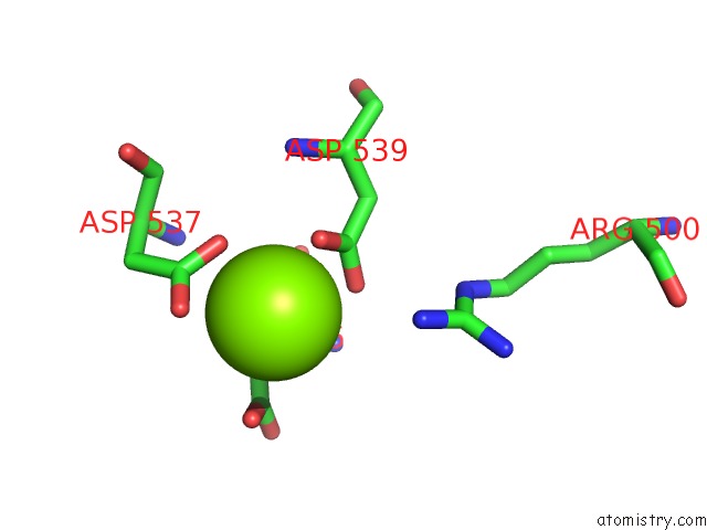
Mono view
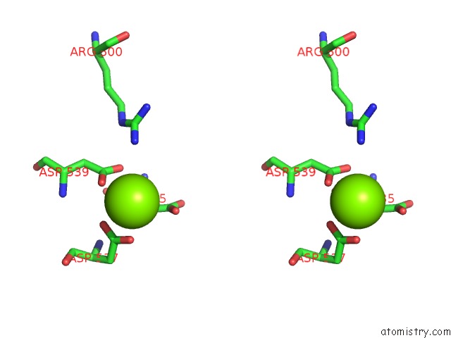
Stereo pair view
Mono view
Stereo pair view
A full contact list of Magnesium with other atoms in the Mg binding
site number 1 of Structure of A Mycobacterium Smegmatis Transcription Initiation Complex with An Upstream-Fork Promoter Fragment within 5.0Å range:
|
Reference:
E.A.Hubin,
M.Lilic,
S.A.Darst,
E.A.Campbell.
Structural Insights Into the Mycobacteria Transcription Initiation Complex From Analysis of X-Ray Crystal Structures. Nat Commun V. 8 16072 2017.
ISSN: ESSN 2041-1723
PubMed: 28703128
DOI: 10.1038/NCOMMS16072
Page generated: Mon Sep 30 06:10:32 2024
ISSN: ESSN 2041-1723
PubMed: 28703128
DOI: 10.1038/NCOMMS16072
Last articles
Zn in 9MJ5Zn in 9HNW
Zn in 9G0L
Zn in 9FNE
Zn in 9DZN
Zn in 9E0I
Zn in 9D32
Zn in 9DAK
Zn in 8ZXC
Zn in 8ZUF