Magnesium »
PDB 5vwa-5w51 »
5vz0 »
Magnesium in PDB 5vz0: Crystal Structure of Lactococcus Lactis Pyruvate Carboxylase G746A Mutant in Complex with Cyclic-Di-Amp
Enzymatic activity of Crystal Structure of Lactococcus Lactis Pyruvate Carboxylase G746A Mutant in Complex with Cyclic-Di-Amp
All present enzymatic activity of Crystal Structure of Lactococcus Lactis Pyruvate Carboxylase G746A Mutant in Complex with Cyclic-Di-Amp:
6.4.1.1;
6.4.1.1;
Protein crystallography data
The structure of Crystal Structure of Lactococcus Lactis Pyruvate Carboxylase G746A Mutant in Complex with Cyclic-Di-Amp, PDB code: 5vz0
was solved by
P.H.Choi,
L.Tong,
with X-Ray Crystallography technique. A brief refinement statistics is given in the table below:
| Resolution Low / High (Å) | 48.55 / 2.00 |
| Space group | P 1 |
| Cell size a, b, c (Å), α, β, γ (°) | 97.155, 130.390, 134.271, 65.99, 88.67, 70.14 |
| R / Rfree (%) | 19.1 / 21.8 |
Other elements in 5vz0:
The structure of Crystal Structure of Lactococcus Lactis Pyruvate Carboxylase G746A Mutant in Complex with Cyclic-Di-Amp also contains other interesting chemical elements:
| Manganese | (Mn) | 4 atoms |
Magnesium Binding Sites:
The binding sites of Magnesium atom in the Crystal Structure of Lactococcus Lactis Pyruvate Carboxylase G746A Mutant in Complex with Cyclic-Di-Amp
(pdb code 5vz0). This binding sites where shown within
5.0 Angstroms radius around Magnesium atom.
In total 3 binding sites of Magnesium where determined in the Crystal Structure of Lactococcus Lactis Pyruvate Carboxylase G746A Mutant in Complex with Cyclic-Di-Amp, PDB code: 5vz0:
Jump to Magnesium binding site number: 1; 2; 3;
In total 3 binding sites of Magnesium where determined in the Crystal Structure of Lactococcus Lactis Pyruvate Carboxylase G746A Mutant in Complex with Cyclic-Di-Amp, PDB code: 5vz0:
Jump to Magnesium binding site number: 1; 2; 3;
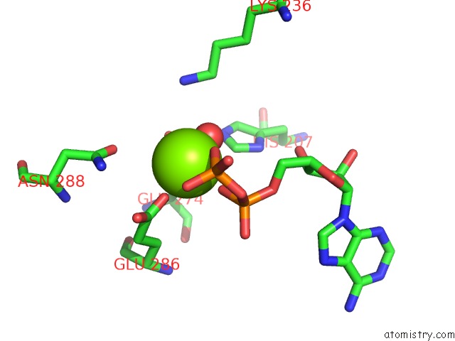
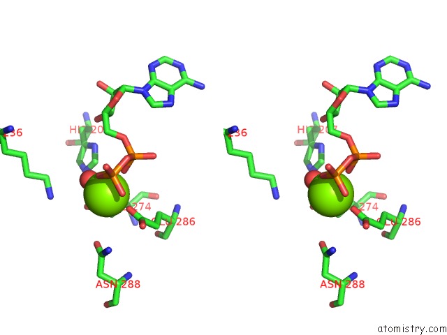
Magnesium binding site 1 out of 3 in 5vz0
Go back to
Magnesium binding site 1 out
of 3 in the Crystal Structure of Lactococcus Lactis Pyruvate Carboxylase G746A Mutant in Complex with Cyclic-Di-Amp
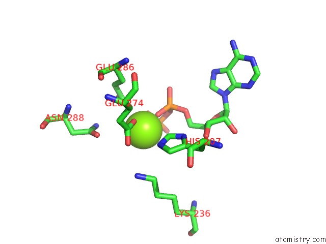
Mono view
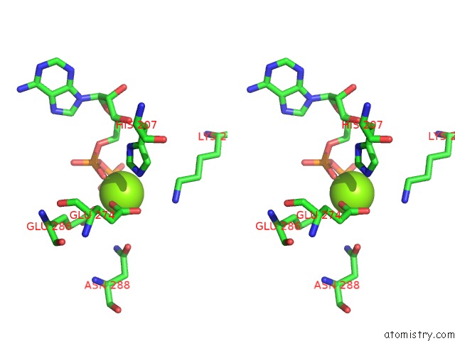
Stereo pair view
Mono view
Stereo pair view
A full contact list of Magnesium with other atoms in the Mg binding
site number 1 of Crystal Structure of Lactococcus Lactis Pyruvate Carboxylase G746A Mutant in Complex with Cyclic-Di-Amp within 5.0Å range:
|
Magnesium binding site 2 out of 3 in 5vz0
Go back to
Magnesium binding site 2 out
of 3 in the Crystal Structure of Lactococcus Lactis Pyruvate Carboxylase G746A Mutant in Complex with Cyclic-Di-Amp
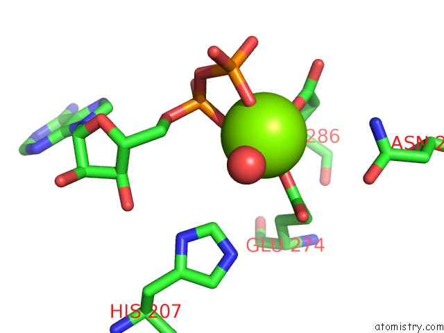
Mono view
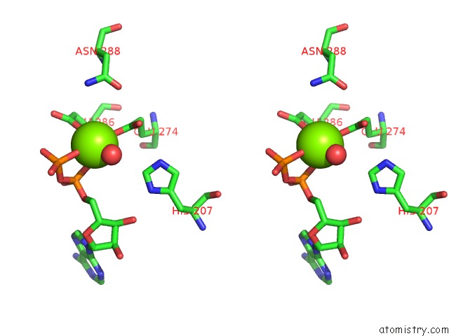
Stereo pair view
Mono view
Stereo pair view
A full contact list of Magnesium with other atoms in the Mg binding
site number 2 of Crystal Structure of Lactococcus Lactis Pyruvate Carboxylase G746A Mutant in Complex with Cyclic-Di-Amp within 5.0Å range:
|
Magnesium binding site 3 out of 3 in 5vz0
Go back to
Magnesium binding site 3 out
of 3 in the Crystal Structure of Lactococcus Lactis Pyruvate Carboxylase G746A Mutant in Complex with Cyclic-Di-Amp
Mono view
Stereo pair view
Mono view
Stereo pair view
A full contact list of Magnesium with other atoms in the Mg binding
site number 3 of Crystal Structure of Lactococcus Lactis Pyruvate Carboxylase G746A Mutant in Complex with Cyclic-Di-Amp within 5.0Å range:
|
Reference:
P.H.Choi,
T.M.N.Vu,
H.T.Pham,
J.J.Woodward,
M.S.Turner,
L.Tong.
Structural and Functional Studies of Pyruvate Carboxylase Regulation By Cyclic Di-Amp in Lactic Acid Bacteria. Proc. Natl. Acad. Sci. V. 114 E7226 2017U.S.A..
ISSN: ESSN 1091-6490
PubMed: 28808024
DOI: 10.1073/PNAS.1704756114
Page generated: Mon Sep 30 06:22:52 2024
ISSN: ESSN 1091-6490
PubMed: 28808024
DOI: 10.1073/PNAS.1704756114
Last articles
Zn in 9MJ5Zn in 9HNW
Zn in 9G0L
Zn in 9FNE
Zn in 9DZN
Zn in 9E0I
Zn in 9D32
Zn in 9DAK
Zn in 8ZXC
Zn in 8ZUF