Magnesium »
PDB 5vw9-5w4u »
5w1z »
Magnesium in PDB 5w1z: Crystal Structure of Inosine-Substituted Decamer Duplex Dna (I4)
Protein crystallography data
The structure of Crystal Structure of Inosine-Substituted Decamer Duplex Dna (I4), PDB code: 5w1z
was solved by
P.S.Pallan,
M.Egli,
with X-Ray Crystallography technique. A brief refinement statistics is given in the table below:
| Resolution Low / High (Å) | 32.24 / 1.55 |
| Space group | P 1 |
| Cell size a, b, c (Å), α, β, γ (°) | 24.884, 32.798, 34.034, 90.25, 107.32, 111.04 |
| R / Rfree (%) | 20.7 / 23.4 |
Other elements in 5w1z:
The structure of Crystal Structure of Inosine-Substituted Decamer Duplex Dna (I4) also contains other interesting chemical elements:
| Bromine | (Br) | 4 atoms |
| Sodium | (Na) | 4 atoms |
Magnesium Binding Sites:
The binding sites of Magnesium atom in the Crystal Structure of Inosine-Substituted Decamer Duplex Dna (I4)
(pdb code 5w1z). This binding sites where shown within
5.0 Angstroms radius around Magnesium atom.
In total 3 binding sites of Magnesium where determined in the Crystal Structure of Inosine-Substituted Decamer Duplex Dna (I4), PDB code: 5w1z:
Jump to Magnesium binding site number: 1; 2; 3;
In total 3 binding sites of Magnesium where determined in the Crystal Structure of Inosine-Substituted Decamer Duplex Dna (I4), PDB code: 5w1z:
Jump to Magnesium binding site number: 1; 2; 3;
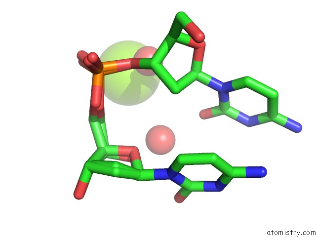
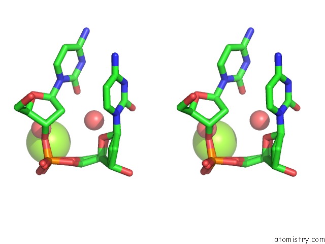
Magnesium binding site 1 out of 3 in 5w1z
Go back to
Magnesium binding site 1 out
of 3 in the Crystal Structure of Inosine-Substituted Decamer Duplex Dna (I4)
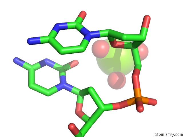
Mono view
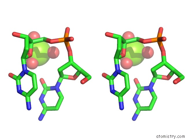
Stereo pair view
Mono view
Stereo pair view
A full contact list of Magnesium with other atoms in the Mg binding
site number 1 of Crystal Structure of Inosine-Substituted Decamer Duplex Dna (I4) within 5.0Å range:
|
Magnesium binding site 2 out of 3 in 5w1z
Go back to
Magnesium binding site 2 out
of 3 in the Crystal Structure of Inosine-Substituted Decamer Duplex Dna (I4)
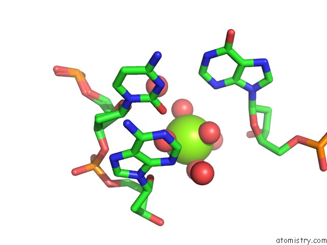
Mono view
Stereo pair view
Mono view
Stereo pair view
A full contact list of Magnesium with other atoms in the Mg binding
site number 2 of Crystal Structure of Inosine-Substituted Decamer Duplex Dna (I4) within 5.0Å range:
|
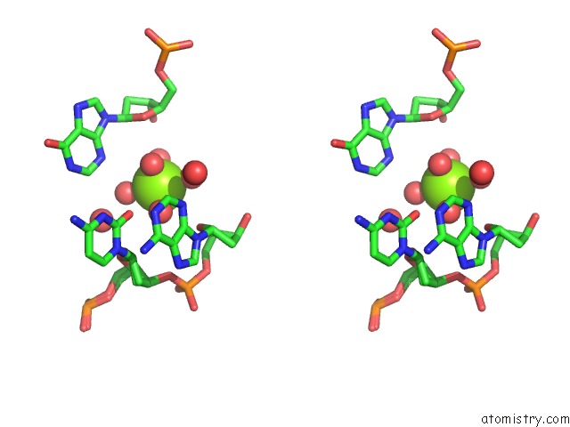
Magnesium binding site 3 out of 3 in 5w1z
Go back to
Magnesium binding site 3 out
of 3 in the Crystal Structure of Inosine-Substituted Decamer Duplex Dna (I4)
Mono view
Stereo pair view
Mono view
Stereo pair view
A full contact list of Magnesium with other atoms in the Mg binding
site number 3 of Crystal Structure of Inosine-Substituted Decamer Duplex Dna (I4) within 5.0Å range:
|
Reference:
J.P.Peters,
E.A.Kowal,
P.S.Pallan,
M.Egli,
L.J.Maher 3Rd..
Comparative Analysis of Inosine-Substituted Duplex Dna By Circular Dichroism and X-Ray Crystallography. J. Biomol. Struct. Dyn. V. 36 2753 2018.
ISSN: ESSN 1538-0254
PubMed: 28818035
DOI: 10.1080/07391102.2017.1369164
Page generated: Mon Sep 30 06:25:55 2024
ISSN: ESSN 1538-0254
PubMed: 28818035
DOI: 10.1080/07391102.2017.1369164
Last articles
Zn in 9J0NZn in 9J0O
Zn in 9J0P
Zn in 9FJX
Zn in 9EKB
Zn in 9C0F
Zn in 9CAH
Zn in 9CH0
Zn in 9CH3
Zn in 9CH1