Magnesium »
PDB 5w5c-5whe »
5wfn »
Magnesium in PDB 5wfn: Revised Model of Leiomodin 2-Mediated Actin Regulation (Alternate Refinement of Pdb 4RWT)
Protein crystallography data
The structure of Revised Model of Leiomodin 2-Mediated Actin Regulation (Alternate Refinement of Pdb 4RWT), PDB code: 5wfn
was solved by
Z.Yurtsever,
M.J.Eck,
R.Dominguez,
with X-Ray Crystallography technique. A brief refinement statistics is given in the table below:
| Resolution Low / High (Å) | 44.25 / 3.00 |
| Space group | P 1 |
| Cell size a, b, c (Å), α, β, γ (°) | 65.350, 65.650, 81.920, 101.29, 90.94, 107.97 |
| R / Rfree (%) | 20.8 / 24.6 |
Magnesium Binding Sites:
The binding sites of Magnesium atom in the Revised Model of Leiomodin 2-Mediated Actin Regulation (Alternate Refinement of Pdb 4RWT)
(pdb code 5wfn). This binding sites where shown within
5.0 Angstroms radius around Magnesium atom.
In total 2 binding sites of Magnesium where determined in the Revised Model of Leiomodin 2-Mediated Actin Regulation (Alternate Refinement of Pdb 4RWT), PDB code: 5wfn:
Jump to Magnesium binding site number: 1; 2;
In total 2 binding sites of Magnesium where determined in the Revised Model of Leiomodin 2-Mediated Actin Regulation (Alternate Refinement of Pdb 4RWT), PDB code: 5wfn:
Jump to Magnesium binding site number: 1; 2;
Magnesium binding site 1 out of 2 in 5wfn
Go back to
Magnesium binding site 1 out
of 2 in the Revised Model of Leiomodin 2-Mediated Actin Regulation (Alternate Refinement of Pdb 4RWT)

Mono view
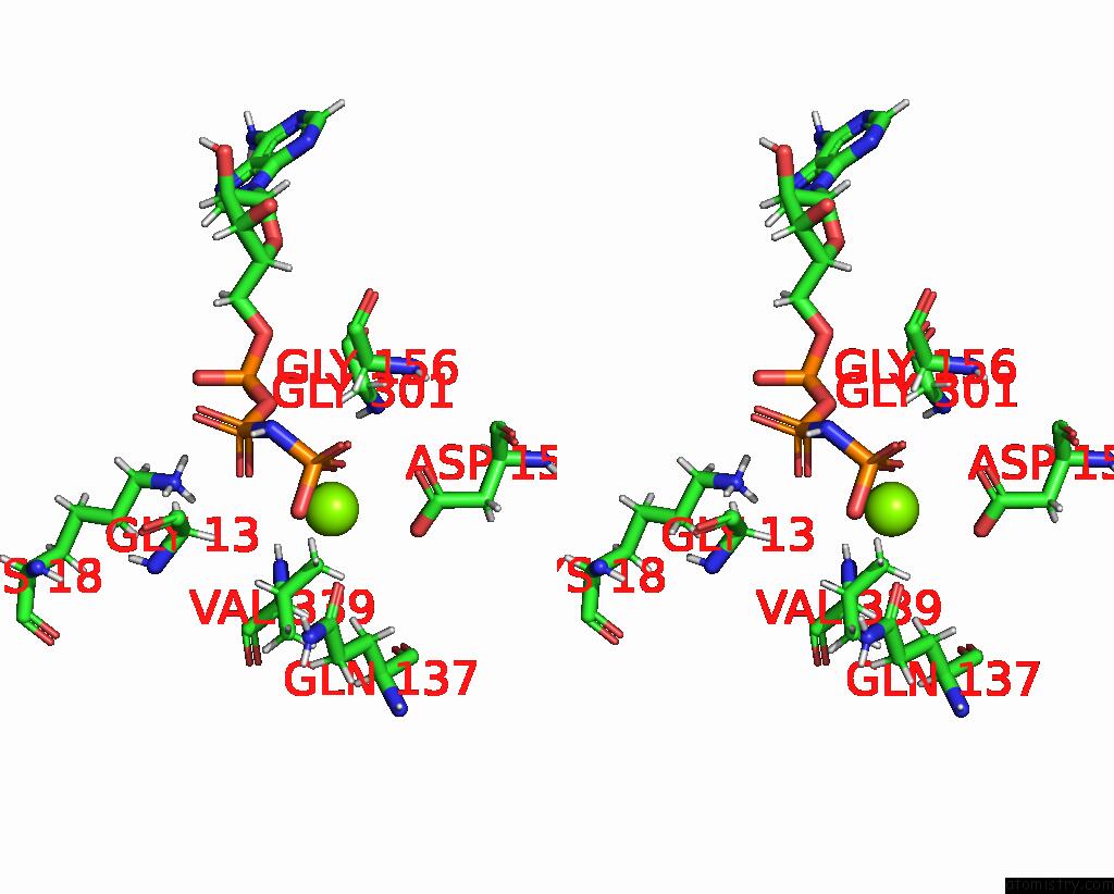
Stereo pair view
Mono view
Stereo pair view
A full contact list of Magnesium with other atoms in the Mg binding
site number 1 of Revised Model of Leiomodin 2-Mediated Actin Regulation (Alternate Refinement of Pdb 4RWT) within 5.0Å range:
|
Magnesium binding site 2 out of 2 in 5wfn
Go back to
Magnesium binding site 2 out
of 2 in the Revised Model of Leiomodin 2-Mediated Actin Regulation (Alternate Refinement of Pdb 4RWT)

Mono view
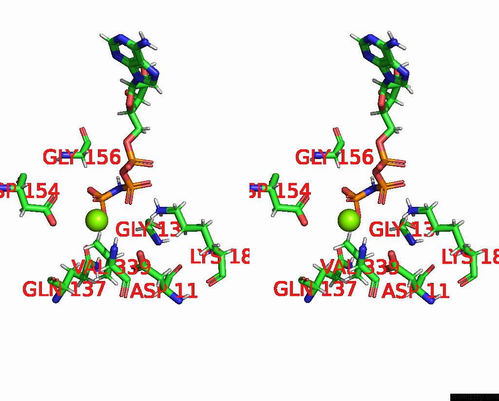
Stereo pair view
Mono view
Stereo pair view
A full contact list of Magnesium with other atoms in the Mg binding
site number 2 of Revised Model of Leiomodin 2-Mediated Actin Regulation (Alternate Refinement of Pdb 4RWT) within 5.0Å range:
|
Reference:
M.Boczkowska,
Z.Yurtsever,
G.Rebowski,
M.J.Eck,
R.Dominguez.
Crystal Structure of Leiomodin 2 in Complex with Actin: A Structural and Functional Reexamination Biophys.J. V. 113 889 2017.
ISSN: ISSN 0006-3495
DOI: 10.1016/J.BPJ.2017.07.007
Page generated: Mon Sep 30 06:33:33 2024
ISSN: ISSN 0006-3495
DOI: 10.1016/J.BPJ.2017.07.007
Last articles
Cl in 3RUTCl in 3RUO
Cl in 3RUN
Cl in 3RU6
Cl in 3ROJ
Cl in 3RTF
Cl in 3RTL
Cl in 3RSP
Cl in 3RT8
Cl in 3RS1