Magnesium »
PDB 5wi5-5wti »
5wqa »
Magnesium in PDB 5wqa: Crystal Structure of PDE4D Catalytic Domain Complexed with Selaginpulvilins K
Enzymatic activity of Crystal Structure of PDE4D Catalytic Domain Complexed with Selaginpulvilins K
All present enzymatic activity of Crystal Structure of PDE4D Catalytic Domain Complexed with Selaginpulvilins K:
3.1.4.53;
3.1.4.53;
Protein crystallography data
The structure of Crystal Structure of PDE4D Catalytic Domain Complexed with Selaginpulvilins K, PDB code: 5wqa
was solved by
Y.Huang,
T.Zhang,
X.Zheng,
S.Yin,
H.B.Luo,
with X-Ray Crystallography technique. A brief refinement statistics is given in the table below:
| Resolution Low / High (Å) | 81.79 / 2.30 |
| Space group | P 21 21 21 |
| Cell size a, b, c (Å), α, β, γ (°) | 57.992, 80.784, 163.575, 90.00, 90.00, 90.00 |
| R / Rfree (%) | 21.7 / 25.2 |
Other elements in 5wqa:
The structure of Crystal Structure of PDE4D Catalytic Domain Complexed with Selaginpulvilins K also contains other interesting chemical elements:
| Zinc | (Zn) | 2 atoms |
Magnesium Binding Sites:
The binding sites of Magnesium atom in the Crystal Structure of PDE4D Catalytic Domain Complexed with Selaginpulvilins K
(pdb code 5wqa). This binding sites where shown within
5.0 Angstroms radius around Magnesium atom.
In total 2 binding sites of Magnesium where determined in the Crystal Structure of PDE4D Catalytic Domain Complexed with Selaginpulvilins K, PDB code: 5wqa:
Jump to Magnesium binding site number: 1; 2;
In total 2 binding sites of Magnesium where determined in the Crystal Structure of PDE4D Catalytic Domain Complexed with Selaginpulvilins K, PDB code: 5wqa:
Jump to Magnesium binding site number: 1; 2;
Magnesium binding site 1 out of 2 in 5wqa
Go back to
Magnesium binding site 1 out
of 2 in the Crystal Structure of PDE4D Catalytic Domain Complexed with Selaginpulvilins K
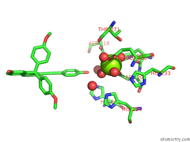
Mono view
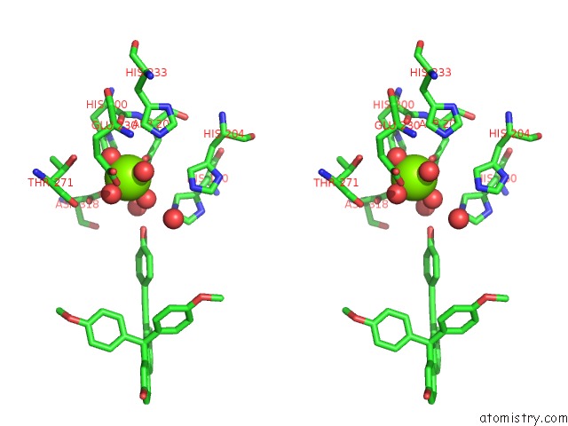
Stereo pair view
Mono view
Stereo pair view
A full contact list of Magnesium with other atoms in the Mg binding
site number 1 of Crystal Structure of PDE4D Catalytic Domain Complexed with Selaginpulvilins K within 5.0Å range:
|
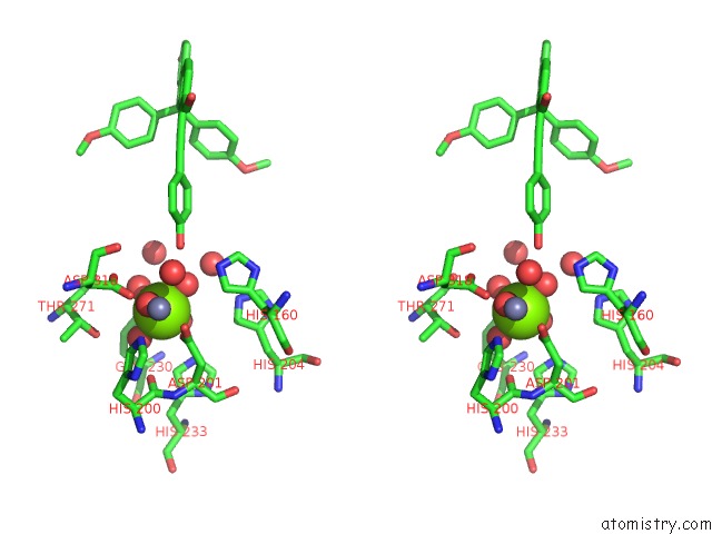
Magnesium binding site 2 out of 2 in 5wqa
Go back to
Magnesium binding site 2 out
of 2 in the Crystal Structure of PDE4D Catalytic Domain Complexed with Selaginpulvilins K
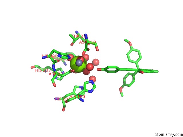
Mono view
Stereo pair view
Mono view
Stereo pair view
A full contact list of Magnesium with other atoms in the Mg binding
site number 2 of Crystal Structure of PDE4D Catalytic Domain Complexed with Selaginpulvilins K within 5.0Å range:
|
Reference:
Y.Huang,
X.Liu,
D.Wu,
G.Tang,
Z.Lai,
X.Zheng,
S.Yin,
H.B.Luo.
The Discovery, Complex Crystal Structure, and Recognition Mechanism of A Novel Natural PDE4 Inhibitor From Selaginella Pulvinata Biochem. Pharmacol. V. 130 51 2017.
ISSN: ISSN 1873-2968
PubMed: 28159622
DOI: 10.1016/J.BCP.2017.01.016
Page generated: Mon Sep 30 06:46:38 2024
ISSN: ISSN 1873-2968
PubMed: 28159622
DOI: 10.1016/J.BCP.2017.01.016
Last articles
Zn in 9MJ5Zn in 9HNW
Zn in 9G0L
Zn in 9FNE
Zn in 9DZN
Zn in 9E0I
Zn in 9D32
Zn in 9DAK
Zn in 8ZXC
Zn in 8ZUF