Magnesium »
PDB 5wti-5x7u »
5x2l »
Magnesium in PDB 5x2l: Crystal Structure of Human Serine Racemase
Enzymatic activity of Crystal Structure of Human Serine Racemase
All present enzymatic activity of Crystal Structure of Human Serine Racemase:
4.3.1.17; 4.3.1.18; 5.1.1.18;
4.3.1.17; 4.3.1.18; 5.1.1.18;
Protein crystallography data
The structure of Crystal Structure of Human Serine Racemase, PDB code: 5x2l
was solved by
T.Obita,
K.Matsumoto,
H.Mori,
N.Toyooka,
M.Mizuguchi,
with X-Ray Crystallography technique. A brief refinement statistics is given in the table below:
| Resolution Low / High (Å) | 46.06 / 1.81 |
| Space group | P 21 21 2 |
| Cell size a, b, c (Å), α, β, γ (°) | 80.120, 112.590, 88.000, 90.00, 90.00, 90.00 |
| R / Rfree (%) | 22 / 25.8 |
Magnesium Binding Sites:
The binding sites of Magnesium atom in the Crystal Structure of Human Serine Racemase
(pdb code 5x2l). This binding sites where shown within
5.0 Angstroms radius around Magnesium atom.
In total 2 binding sites of Magnesium where determined in the Crystal Structure of Human Serine Racemase, PDB code: 5x2l:
Jump to Magnesium binding site number: 1; 2;
In total 2 binding sites of Magnesium where determined in the Crystal Structure of Human Serine Racemase, PDB code: 5x2l:
Jump to Magnesium binding site number: 1; 2;
Magnesium binding site 1 out of 2 in 5x2l
Go back to
Magnesium binding site 1 out
of 2 in the Crystal Structure of Human Serine Racemase
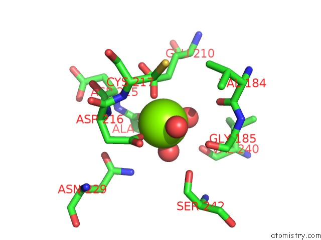
Mono view
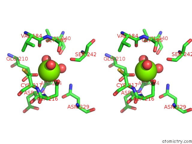
Stereo pair view
Mono view
Stereo pair view
A full contact list of Magnesium with other atoms in the Mg binding
site number 1 of Crystal Structure of Human Serine Racemase within 5.0Å range:
|
Magnesium binding site 2 out of 2 in 5x2l
Go back to
Magnesium binding site 2 out
of 2 in the Crystal Structure of Human Serine Racemase
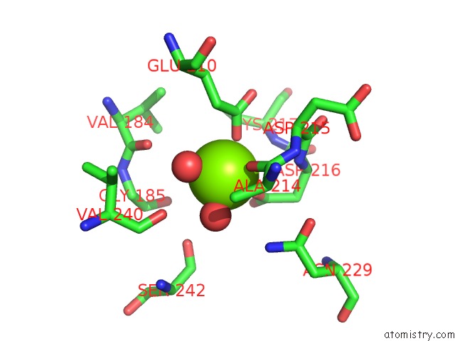
Mono view
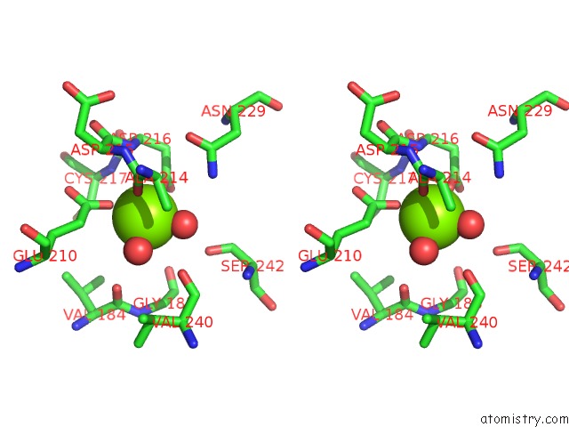
Stereo pair view
Mono view
Stereo pair view
A full contact list of Magnesium with other atoms in the Mg binding
site number 2 of Crystal Structure of Human Serine Racemase within 5.0Å range:
|
Reference:
S.Takahara,
K.Nakagawa,
T.Uchiyama,
T.Yoshida,
K.Matsumoto,
Y.Kawasumi,
M.Mizuguchi,
T.Obita,
Y.Watanabe,
D.Hayakawa,
H.Gouda,
H.Mori,
N.Toyooka.
Design, Synthesis, and Evaluation of Novel Inhibitors For Wild-Type Human Serine Racemase. Bioorg. Med. Chem. Lett. 2017.
ISSN: ESSN 1464-3405
PubMed: 29277459
DOI: 10.1016/J.BMCL.2017.12.021
Page generated: Mon Sep 30 09:00:29 2024
ISSN: ESSN 1464-3405
PubMed: 29277459
DOI: 10.1016/J.BMCL.2017.12.021
Last articles
Zn in 9JYWZn in 9IR4
Zn in 9IR3
Zn in 9GMX
Zn in 9GMW
Zn in 9JEJ
Zn in 9ERF
Zn in 9ERE
Zn in 9EGV
Zn in 9EGW