Magnesium »
PDB 5wti-5x7u »
5x6i »
Magnesium in PDB 5x6i: Crystal Structure of B. Subtilis Adenylate Kinase Variant
Enzymatic activity of Crystal Structure of B. Subtilis Adenylate Kinase Variant
All present enzymatic activity of Crystal Structure of B. Subtilis Adenylate Kinase Variant:
2.7.4.3;
2.7.4.3;
Protein crystallography data
The structure of Crystal Structure of B. Subtilis Adenylate Kinase Variant, PDB code: 5x6i
was solved by
S.Moon,
E.Bae,
with X-Ray Crystallography technique. A brief refinement statistics is given in the table below:
| Resolution Low / High (Å) | 50.00 / 2.00 |
| Space group | P 21 21 21 |
| Cell size a, b, c (Å), α, β, γ (°) | 43.739, 44.180, 100.595, 90.00, 90.00, 90.00 |
| R / Rfree (%) | 17.9 / 25.9 |
Other elements in 5x6i:
The structure of Crystal Structure of B. Subtilis Adenylate Kinase Variant also contains other interesting chemical elements:
| Zinc | (Zn) | 1 atom |
| Calcium | (Ca) | 2 atoms |
Magnesium Binding Sites:
The binding sites of Magnesium atom in the Crystal Structure of B. Subtilis Adenylate Kinase Variant
(pdb code 5x6i). This binding sites where shown within
5.0 Angstroms radius around Magnesium atom.
In total only one binding site of Magnesium was determined in the Crystal Structure of B. Subtilis Adenylate Kinase Variant, PDB code: 5x6i:
In total only one binding site of Magnesium was determined in the Crystal Structure of B. Subtilis Adenylate Kinase Variant, PDB code: 5x6i:
Magnesium binding site 1 out of 1 in 5x6i
Go back to
Magnesium binding site 1 out
of 1 in the Crystal Structure of B. Subtilis Adenylate Kinase Variant
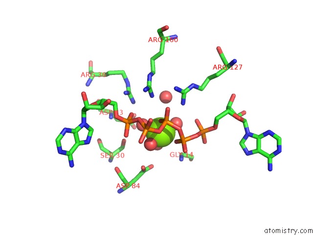
Mono view
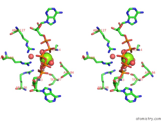
Stereo pair view
Mono view
Stereo pair view
A full contact list of Magnesium with other atoms in the Mg binding
site number 1 of Crystal Structure of B. Subtilis Adenylate Kinase Variant within 5.0Å range:
|
Reference:
S.Moon,
J.Kim,
J.Koo,
E.Bae.
Structural and Mutational Analyses of Psychrophilic and Mesophilic Adenylate Kinases Highlight the Role of Hydrophobic Interactions in Protein Thermal Stability. Struct Dyn. V. 6 24702 2019.
ISSN: ESSN 2329-7778
PubMed: 31111079
DOI: 10.1063/1.5089707
Page generated: Mon Sep 30 09:02:10 2024
ISSN: ESSN 2329-7778
PubMed: 31111079
DOI: 10.1063/1.5089707
Last articles
Zn in 9J0NZn in 9J0O
Zn in 9J0P
Zn in 9FJX
Zn in 9EKB
Zn in 9C0F
Zn in 9CAH
Zn in 9CH0
Zn in 9CH3
Zn in 9CH1