Magnesium »
PDB 5wu3-5x86 »
5x7r »
Magnesium in PDB 5x7r: Crystal Structure of Paenibacillus Sp. 598K Alpha-1,6- Glucosyltransferase Complexed with Isomaltohexaose
Enzymatic activity of Crystal Structure of Paenibacillus Sp. 598K Alpha-1,6- Glucosyltransferase Complexed with Isomaltohexaose
All present enzymatic activity of Crystal Structure of Paenibacillus Sp. 598K Alpha-1,6- Glucosyltransferase Complexed with Isomaltohexaose:
3.2.1.20;
3.2.1.20;
Protein crystallography data
The structure of Crystal Structure of Paenibacillus Sp. 598K Alpha-1,6- Glucosyltransferase Complexed with Isomaltohexaose, PDB code: 5x7r
was solved by
Z.Fujimoto,
N.Kishine,
N.Suzuki,
M.Momma,
H.Ichinose,
A.Kimura,
K.Funane,
with X-Ray Crystallography technique. A brief refinement statistics is given in the table below:
| Resolution Low / High (Å) | 152.46 / 1.95 |
| Space group | C 2 2 21 |
| Cell size a, b, c (Å), α, β, γ (°) | 184.333, 271.260, 133.928, 90.00, 90.00, 90.00 |
| R / Rfree (%) | 17.8 / 20.8 |
Other elements in 5x7r:
The structure of Crystal Structure of Paenibacillus Sp. 598K Alpha-1,6- Glucosyltransferase Complexed with Isomaltohexaose also contains other interesting chemical elements:
| Nickel | (Ni) | 4 atoms |
| Calcium | (Ca) | 6 atoms |
Magnesium Binding Sites:
Pages:
>>> Page 1 <<< Page 2, Binding sites: 11 - 20;Binding sites:
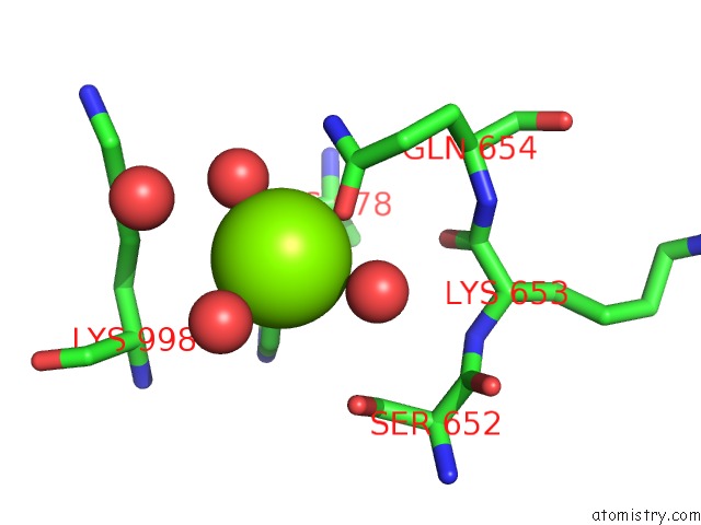
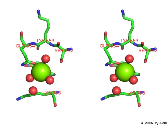
The binding sites of Magnesium atom in the Crystal Structure of Paenibacillus Sp. 598K Alpha-1,6- Glucosyltransferase Complexed with Isomaltohexaose (pdb code 5x7r). This binding sites where shown within 5.0 Angstroms radius around Magnesium atom.In total 20 binding sites of Magnesium where determined in the Crystal Structure of Paenibacillus Sp. 598K Alpha-1,6- Glucosyltransferase Complexed with Isomaltohexaose, PDB code: 5x7r:
Jump to Magnesium binding site number: 1; 2; 3; 4; 5; 6; 7; 8; 9; 10;
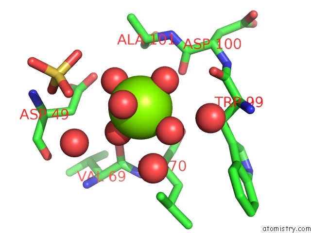
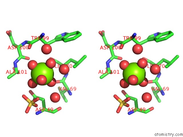
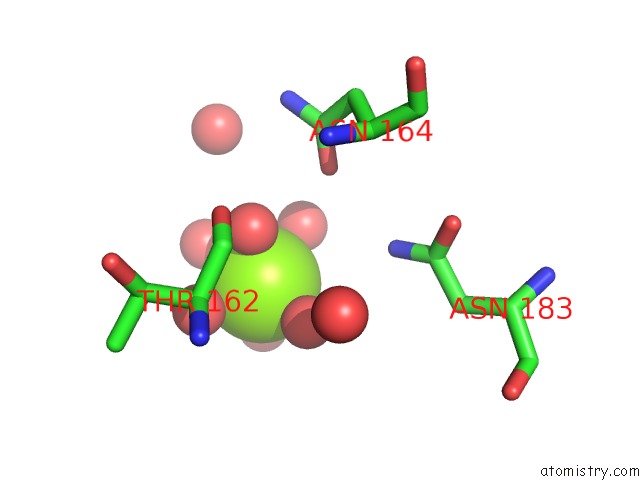
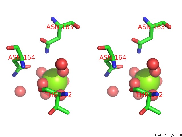
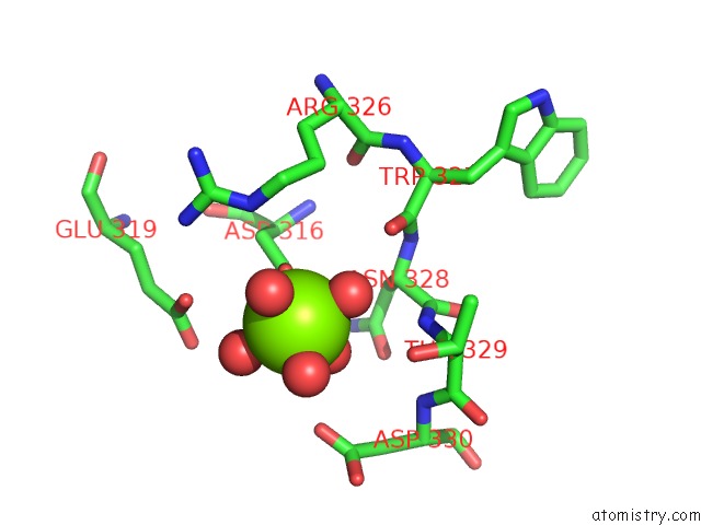
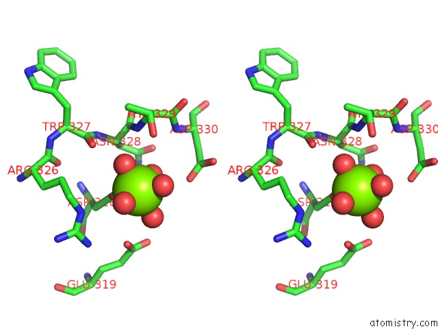
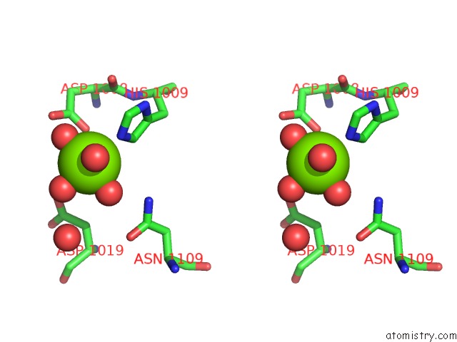
Magnesium binding site 1 out of 20 in 5x7r
Go back to
Magnesium binding site 1 out
of 20 in the Crystal Structure of Paenibacillus Sp. 598K Alpha-1,6- Glucosyltransferase Complexed with Isomaltohexaose
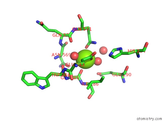
Mono view
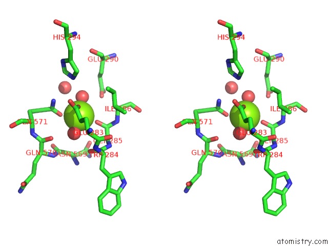
Stereo pair view
Mono view
Stereo pair view
A full contact list of Magnesium with other atoms in the Mg binding
site number 1 of Crystal Structure of Paenibacillus Sp. 598K Alpha-1,6- Glucosyltransferase Complexed with Isomaltohexaose within 5.0Å range:
|
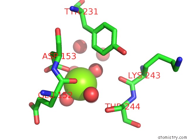
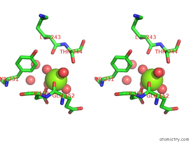
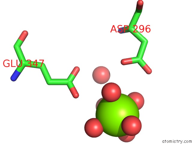
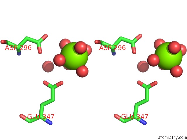
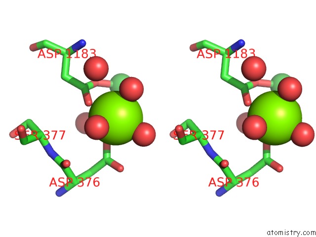
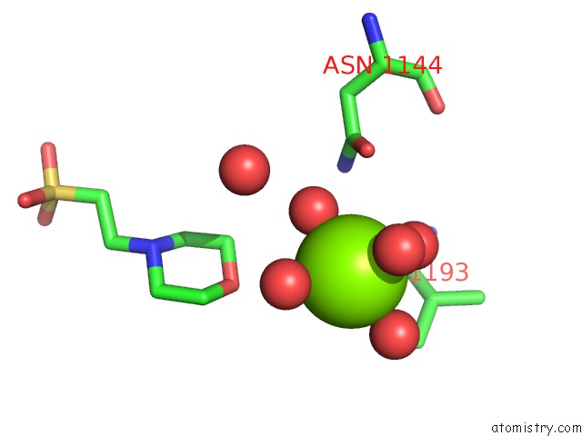
Magnesium binding site 2 out of 20 in 5x7r
Go back to
Magnesium binding site 2 out
of 20 in the Crystal Structure of Paenibacillus Sp. 598K Alpha-1,6- Glucosyltransferase Complexed with Isomaltohexaose
Mono view
Stereo pair view
Mono view
Stereo pair view
A full contact list of Magnesium with other atoms in the Mg binding
site number 2 of Crystal Structure of Paenibacillus Sp. 598K Alpha-1,6- Glucosyltransferase Complexed with Isomaltohexaose within 5.0Å range:
|
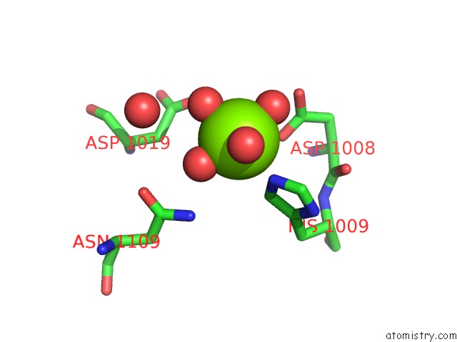
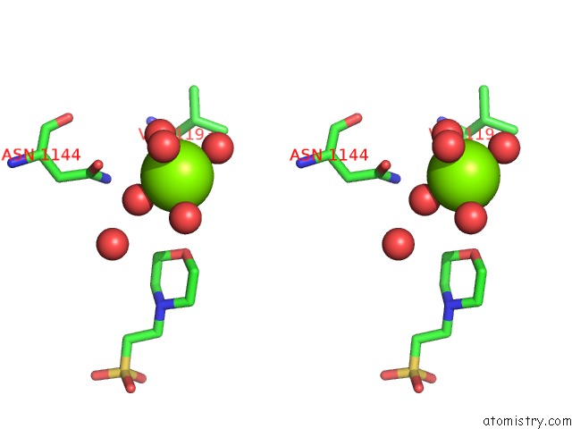
Magnesium binding site 3 out of 20 in 5x7r
Go back to
Magnesium binding site 3 out
of 20 in the Crystal Structure of Paenibacillus Sp. 598K Alpha-1,6- Glucosyltransferase Complexed with Isomaltohexaose
Mono view
Stereo pair view
Mono view
Stereo pair view
A full contact list of Magnesium with other atoms in the Mg binding
site number 3 of Crystal Structure of Paenibacillus Sp. 598K Alpha-1,6- Glucosyltransferase Complexed with Isomaltohexaose within 5.0Å range:
|
Magnesium binding site 4 out of 20 in 5x7r
Go back to
Magnesium binding site 4 out
of 20 in the Crystal Structure of Paenibacillus Sp. 598K Alpha-1,6- Glucosyltransferase Complexed with Isomaltohexaose
Mono view
Stereo pair view
Mono view
Stereo pair view
A full contact list of Magnesium with other atoms in the Mg binding
site number 4 of Crystal Structure of Paenibacillus Sp. 598K Alpha-1,6- Glucosyltransferase Complexed with Isomaltohexaose within 5.0Å range:
|
Magnesium binding site 5 out of 20 in 5x7r
Go back to
Magnesium binding site 5 out
of 20 in the Crystal Structure of Paenibacillus Sp. 598K Alpha-1,6- Glucosyltransferase Complexed with Isomaltohexaose
Mono view
Stereo pair view
Mono view
Stereo pair view
A full contact list of Magnesium with other atoms in the Mg binding
site number 5 of Crystal Structure of Paenibacillus Sp. 598K Alpha-1,6- Glucosyltransferase Complexed with Isomaltohexaose within 5.0Å range:
|
Magnesium binding site 6 out of 20 in 5x7r
Go back to
Magnesium binding site 6 out
of 20 in the Crystal Structure of Paenibacillus Sp. 598K Alpha-1,6- Glucosyltransferase Complexed with Isomaltohexaose
Mono view
Stereo pair view
Mono view
Stereo pair view
A full contact list of Magnesium with other atoms in the Mg binding
site number 6 of Crystal Structure of Paenibacillus Sp. 598K Alpha-1,6- Glucosyltransferase Complexed with Isomaltohexaose within 5.0Å range:
|
Magnesium binding site 7 out of 20 in 5x7r
Go back to
Magnesium binding site 7 out
of 20 in the Crystal Structure of Paenibacillus Sp. 598K Alpha-1,6- Glucosyltransferase Complexed with Isomaltohexaose
Mono view
Stereo pair view
Mono view
Stereo pair view
A full contact list of Magnesium with other atoms in the Mg binding
site number 7 of Crystal Structure of Paenibacillus Sp. 598K Alpha-1,6- Glucosyltransferase Complexed with Isomaltohexaose within 5.0Å range:
|
Magnesium binding site 8 out of 20 in 5x7r
Go back to
Magnesium binding site 8 out
of 20 in the Crystal Structure of Paenibacillus Sp. 598K Alpha-1,6- Glucosyltransferase Complexed with Isomaltohexaose

Mono view
Stereo pair view
Mono view
Stereo pair view
A full contact list of Magnesium with other atoms in the Mg binding
site number 8 of Crystal Structure of Paenibacillus Sp. 598K Alpha-1,6- Glucosyltransferase Complexed with Isomaltohexaose within 5.0Å range:
|
Magnesium binding site 9 out of 20 in 5x7r
Go back to
Magnesium binding site 9 out
of 20 in the Crystal Structure of Paenibacillus Sp. 598K Alpha-1,6- Glucosyltransferase Complexed with Isomaltohexaose
Mono view
Stereo pair view
Mono view
Stereo pair view
A full contact list of Magnesium with other atoms in the Mg binding
site number 9 of Crystal Structure of Paenibacillus Sp. 598K Alpha-1,6- Glucosyltransferase Complexed with Isomaltohexaose within 5.0Å range:
|
Magnesium binding site 10 out of 20 in 5x7r
Go back to
Magnesium binding site 10 out
of 20 in the Crystal Structure of Paenibacillus Sp. 598K Alpha-1,6- Glucosyltransferase Complexed with Isomaltohexaose
Mono view
Stereo pair view
Mono view
Stereo pair view
A full contact list of Magnesium with other atoms in the Mg binding
site number 10 of Crystal Structure of Paenibacillus Sp. 598K Alpha-1,6- Glucosyltransferase Complexed with Isomaltohexaose within 5.0Å range:
|
Reference:
Z.Fujimoto,
N.Suzuki,
N.Kishine,
H.Ichinose,
M.Momma,
A.Kimura,
K.Funane.
Carbohydrate-Binding Architecture of the Multi-Modular Alpha-1,6-Glucosyltransferase From Paenibacillus Sp. 598K, Which Produces Alpha-1,6-Glucosyl-Alpha-Glucosaccharides From Starch Biochem. J. V. 474 2763 2017.
ISSN: ESSN 1470-8728
PubMed: 28698247
DOI: 10.1042/BCJ20170152
Page generated: Mon Sep 30 09:03:06 2024
ISSN: ESSN 1470-8728
PubMed: 28698247
DOI: 10.1042/BCJ20170152
Last articles
Zn in 9MJ5Zn in 9HNW
Zn in 9G0L
Zn in 9FNE
Zn in 9DZN
Zn in 9E0I
Zn in 9D32
Zn in 9DAK
Zn in 8ZXC
Zn in 8ZUF