Magnesium »
PDB 5x86-5xf5 »
5xc5 »
Magnesium in PDB 5xc5: Crystal Structure of Acanthamoeba Polyphaga Mimivirus Rab Gtpase in Complex with Gtp
Protein crystallography data
The structure of Crystal Structure of Acanthamoeba Polyphaga Mimivirus Rab Gtpase in Complex with Gtp, PDB code: 5xc5
was solved by
B.Ku,
J.A.You,
S.J.Kim,
with X-Ray Crystallography technique. A brief refinement statistics is given in the table below:
| Resolution Low / High (Å) | 31.16 / 1.40 |
| Space group | P 21 21 21 |
| Cell size a, b, c (Å), α, β, γ (°) | 32.924, 53.724, 96.482, 90.00, 90.00, 90.00 |
| R / Rfree (%) | 17.4 / 19 |
Magnesium Binding Sites:
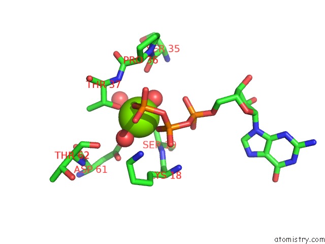
The binding sites of Magnesium atom in the Crystal Structure of Acanthamoeba Polyphaga Mimivirus Rab Gtpase in Complex with Gtp
(pdb code 5xc5). This binding sites where shown within
5.0 Angstroms radius around Magnesium atom.
In total only one binding site of Magnesium was determined in the Crystal Structure of Acanthamoeba Polyphaga Mimivirus Rab Gtpase in Complex with Gtp, PDB code: 5xc5:
In total only one binding site of Magnesium was determined in the Crystal Structure of Acanthamoeba Polyphaga Mimivirus Rab Gtpase in Complex with Gtp, PDB code: 5xc5:
Magnesium binding site 1 out of 1 in 5xc5
Go back to
Magnesium binding site 1 out
of 1 in the Crystal Structure of Acanthamoeba Polyphaga Mimivirus Rab Gtpase in Complex with Gtp
Mono view

Stereo pair view
Mono view
Stereo pair view
A full contact list of Magnesium with other atoms in the Mg binding
site number 1 of Crystal Structure of Acanthamoeba Polyphaga Mimivirus Rab Gtpase in Complex with Gtp within 5.0Å range:
|
Reference:
B.Ku,
J.A.You,
K.J.Oh,
H.Y.Yun,
H.S.Lee,
H.C.Shin,
J.Jung,
Y.B.Shin,
S.J.Kim.
Crystal Structures of Two Forms of the Acanthamoeba Polyphaga Mimivirus Rab Gtpase Arch. Virol. V. 162 3407 2017.
ISSN: ISSN 1432-8798
PubMed: 28779233
DOI: 10.1007/S00705-017-3510-2
Page generated: Mon Sep 30 09:16:56 2024
ISSN: ISSN 1432-8798
PubMed: 28779233
DOI: 10.1007/S00705-017-3510-2
Last articles
Zn in 9J0NZn in 9J0O
Zn in 9J0P
Zn in 9FJX
Zn in 9EKB
Zn in 9C0F
Zn in 9CAH
Zn in 9CH0
Zn in 9CH3
Zn in 9CH1