Magnesium »
PDB 5x86-5xf5 »
5xd7 »
Magnesium in PDB 5xd7: Crystal Structure Analysis of 3,6-Anhydro-L-Galactonate Cycloisomerase
Enzymatic activity of Crystal Structure Analysis of 3,6-Anhydro-L-Galactonate Cycloisomerase
All present enzymatic activity of Crystal Structure Analysis of 3,6-Anhydro-L-Galactonate Cycloisomerase:
5.5.1.25;
5.5.1.25;
Protein crystallography data
The structure of Crystal Structure Analysis of 3,6-Anhydro-L-Galactonate Cycloisomerase, PDB code: 5xd7
was solved by
S.Lee,
I.-G.Choi,
H.-Y.Kim,
with X-Ray Crystallography technique. A brief refinement statistics is given in the table below:
| Resolution Low / High (Å) | 37.68 / 2.20 |
| Space group | P 41 21 2 |
| Cell size a, b, c (Å), α, β, γ (°) | 87.336, 87.336, 143.069, 90.00, 90.00, 90.00 |
| R / Rfree (%) | 18.8 / 23.8 |
Magnesium Binding Sites:
The binding sites of Magnesium atom in the Crystal Structure Analysis of 3,6-Anhydro-L-Galactonate Cycloisomerase
(pdb code 5xd7). This binding sites where shown within
5.0 Angstroms radius around Magnesium atom.
In total 2 binding sites of Magnesium where determined in the Crystal Structure Analysis of 3,6-Anhydro-L-Galactonate Cycloisomerase, PDB code: 5xd7:
Jump to Magnesium binding site number: 1; 2;
In total 2 binding sites of Magnesium where determined in the Crystal Structure Analysis of 3,6-Anhydro-L-Galactonate Cycloisomerase, PDB code: 5xd7:
Jump to Magnesium binding site number: 1; 2;
Magnesium binding site 1 out of 2 in 5xd7
Go back to
Magnesium binding site 1 out
of 2 in the Crystal Structure Analysis of 3,6-Anhydro-L-Galactonate Cycloisomerase

Mono view
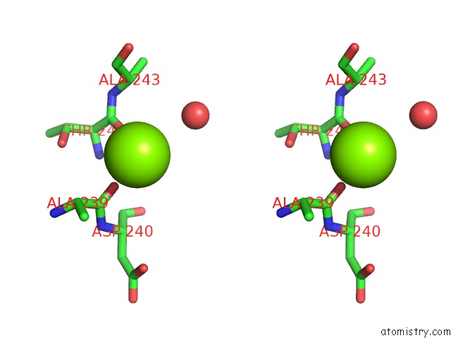
Stereo pair view
Mono view
Stereo pair view
A full contact list of Magnesium with other atoms in the Mg binding
site number 1 of Crystal Structure Analysis of 3,6-Anhydro-L-Galactonate Cycloisomerase within 5.0Å range:
|
Magnesium binding site 2 out of 2 in 5xd7
Go back to
Magnesium binding site 2 out
of 2 in the Crystal Structure Analysis of 3,6-Anhydro-L-Galactonate Cycloisomerase

Mono view

Stereo pair view
Mono view
Stereo pair view
A full contact list of Magnesium with other atoms in the Mg binding
site number 2 of Crystal Structure Analysis of 3,6-Anhydro-L-Galactonate Cycloisomerase within 5.0Å range:
|
Reference:
S.Lee,
K.H.Kim,
H.-Y.Kim,
I.-G.Choi.
Crystal Structure Analysis of 3,6-Anhydro-L-Galactonate Cycloisomerase Suggests Emergence of Novel Substrate Specificity in the Enolase Superfamily. Biochem. Biophys. Res. V. 491 217 2017COMMUN..
ISSN: ESSN 1090-2104
PubMed: 28716734
DOI: 10.1016/J.BBRC.2017.07.080
Page generated: Mon Sep 30 09:18:07 2024
ISSN: ESSN 1090-2104
PubMed: 28716734
DOI: 10.1016/J.BBRC.2017.07.080
Last articles
Zn in 9J0NZn in 9J0O
Zn in 9J0P
Zn in 9FJX
Zn in 9EKB
Zn in 9C0F
Zn in 9CAH
Zn in 9CH0
Zn in 9CH3
Zn in 9CH1