Magnesium »
PDB 5x8b-5xf9 »
5xd8 »
Magnesium in PDB 5xd8: Crystal Structure Analysis of 3,6-Anhydro-L-Galactonate Cycloisomerase
Enzymatic activity of Crystal Structure Analysis of 3,6-Anhydro-L-Galactonate Cycloisomerase
All present enzymatic activity of Crystal Structure Analysis of 3,6-Anhydro-L-Galactonate Cycloisomerase:
5.5.1.25;
5.5.1.25;
Protein crystallography data
The structure of Crystal Structure Analysis of 3,6-Anhydro-L-Galactonate Cycloisomerase, PDB code: 5xd8
was solved by
S.Lee,
I.-G.Choi,
H.-Y.Kim,
with X-Ray Crystallography technique. A brief refinement statistics is given in the table below:
| Resolution Low / High (Å) | 39.14 / 2.51 |
| Space group | P 21 21 21 |
| Cell size a, b, c (Å), α, β, γ (°) | 84.487, 90.350, 104.019, 90.00, 90.00, 90.00 |
| R / Rfree (%) | 20.9 / 27.7 |
Magnesium Binding Sites:
The binding sites of Magnesium atom in the Crystal Structure Analysis of 3,6-Anhydro-L-Galactonate Cycloisomerase
(pdb code 5xd8). This binding sites where shown within
5.0 Angstroms radius around Magnesium atom.
In total 2 binding sites of Magnesium where determined in the Crystal Structure Analysis of 3,6-Anhydro-L-Galactonate Cycloisomerase, PDB code: 5xd8:
Jump to Magnesium binding site number: 1; 2;
In total 2 binding sites of Magnesium where determined in the Crystal Structure Analysis of 3,6-Anhydro-L-Galactonate Cycloisomerase, PDB code: 5xd8:
Jump to Magnesium binding site number: 1; 2;
Magnesium binding site 1 out of 2 in 5xd8
Go back to
Magnesium binding site 1 out
of 2 in the Crystal Structure Analysis of 3,6-Anhydro-L-Galactonate Cycloisomerase

Mono view
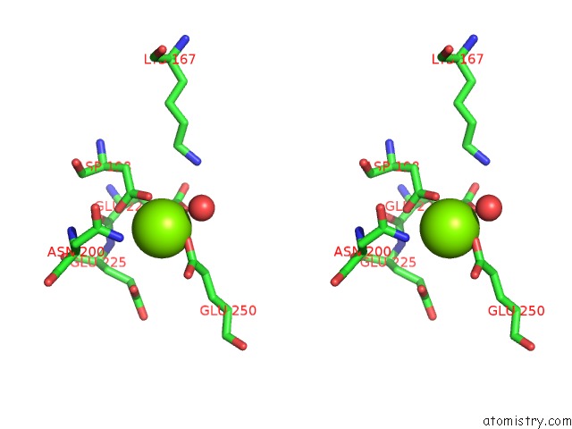
Stereo pair view
Mono view
Stereo pair view
A full contact list of Magnesium with other atoms in the Mg binding
site number 1 of Crystal Structure Analysis of 3,6-Anhydro-L-Galactonate Cycloisomerase within 5.0Å range:
|
Magnesium binding site 2 out of 2 in 5xd8
Go back to
Magnesium binding site 2 out
of 2 in the Crystal Structure Analysis of 3,6-Anhydro-L-Galactonate Cycloisomerase
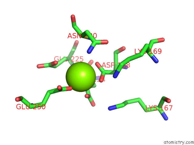
Mono view
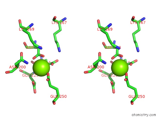
Stereo pair view
Mono view
Stereo pair view
A full contact list of Magnesium with other atoms in the Mg binding
site number 2 of Crystal Structure Analysis of 3,6-Anhydro-L-Galactonate Cycloisomerase within 5.0Å range:
|
Reference:
S.Lee,
K.H.Kim,
H.-Y.Kim,
I.-G.Choi.
Crystal Structure Analysis of 3,6-Anhydro-L-Galactonate Cycloisomerase Suggests Emergence of Novel Substrate Specificity in the Enolase Superfamily Biochem. Biophys. Res. V. 491 217 2017COMMUN..
ISSN: ESSN 1090-2104
PubMed: 28716734
DOI: 10.1016/J.BBRC.2017.07.080
Page generated: Tue Aug 12 23:30:47 2025
ISSN: ESSN 1090-2104
PubMed: 28716734
DOI: 10.1016/J.BBRC.2017.07.080
Last articles
Mg in 6KD7Mg in 6KCM
Mg in 6KCK
Mg in 6KCL
Mg in 6KCC
Mg in 6KCA
Mg in 6KC0
Mg in 6KBZ
Mg in 6KAL
Mg in 6KAK