Magnesium »
PDB 5xf9-5xlf »
5xfe »
Magnesium in PDB 5xfe: Luciferin-Regenerating Enzyme Solved By Sad Using Xfel (Refined Against 11,000 Patterns)
Protein crystallography data
The structure of Luciferin-Regenerating Enzyme Solved By Sad Using Xfel (Refined Against 11,000 Patterns), PDB code: 5xfe
was solved by
K.Yamashita,
D.Pan,
T.Okuda,
T.Murai,
A.Kodan,
T.Yamaguchi,
K.Gomi,
N.Kajiyama,
H.Kato,
H.Ago,
M.Yamamoto,
T.Nakatsu,
with X-Ray Crystallography technique. A brief refinement statistics is given in the table below:
| Resolution Low / High (Å) | 24.57 / 1.50 |
| Space group | P 21 21 21 |
| Cell size a, b, c (Å), α, β, γ (°) | 47.900, 77.030, 84.530, 90.00, 90.00, 90.00 |
| R / Rfree (%) | 19.2 / 21.3 |
Other elements in 5xfe:
The structure of Luciferin-Regenerating Enzyme Solved By Sad Using Xfel (Refined Against 11,000 Patterns) also contains other interesting chemical elements:
| Mercury | (Hg) | 2 atoms |
Magnesium Binding Sites:
The binding sites of Magnesium atom in the Luciferin-Regenerating Enzyme Solved By Sad Using Xfel (Refined Against 11,000 Patterns)
(pdb code 5xfe). This binding sites where shown within
5.0 Angstroms radius around Magnesium atom.
In total only one binding site of Magnesium was determined in the Luciferin-Regenerating Enzyme Solved By Sad Using Xfel (Refined Against 11,000 Patterns), PDB code: 5xfe:
In total only one binding site of Magnesium was determined in the Luciferin-Regenerating Enzyme Solved By Sad Using Xfel (Refined Against 11,000 Patterns), PDB code: 5xfe:
Magnesium binding site 1 out of 1 in 5xfe
Go back to
Magnesium binding site 1 out
of 1 in the Luciferin-Regenerating Enzyme Solved By Sad Using Xfel (Refined Against 11,000 Patterns)
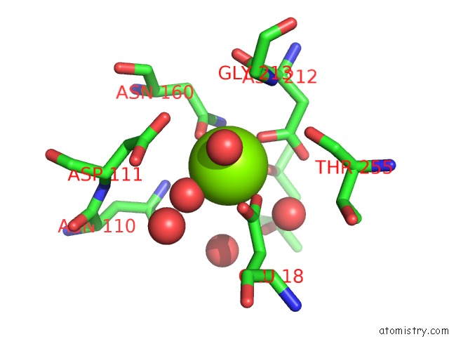
Mono view
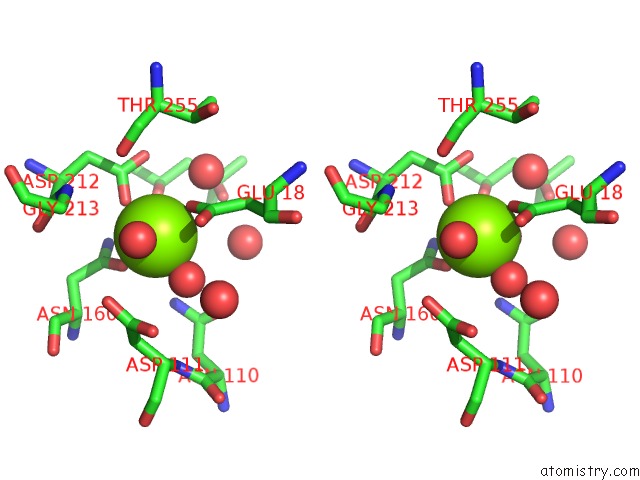
Stereo pair view
Mono view
Stereo pair view
A full contact list of Magnesium with other atoms in the Mg binding
site number 1 of Luciferin-Regenerating Enzyme Solved By Sad Using Xfel (Refined Against 11,000 Patterns) within 5.0Å range:
|
Reference:
K.Yamashita,
N.Kuwabara,
T.Nakane,
T.Murai,
E.Mizohata,
M.Sugahara,
D.Pan,
T.Masuda,
M.Suzuki,
T.Sato,
A.Kodan,
T.Yamaguchi,
E.Nango,
T.Tanaka,
K.Tono,
Y.Joti,
T.Kameshima,
T.Hatsui,
M.Yabashi,
H.Manya,
T.Endo,
R.Kato,
T.Senda,
H.Kato,
S.Iwata,
H.Ago,
M.Yamamoto,
F.Yumoto,
T.Nakatsu.
Experimental Phase Determination with Selenomethionine or Mercury-Derivatization in Serial Femtosecond Crystallography Iucrj V. 4 639 2017.
ISSN: ESSN 2052-2525
PubMed: 28989719
DOI: 10.1107/S2052252517008557
Page generated: Mon Sep 30 09:21:46 2024
ISSN: ESSN 2052-2525
PubMed: 28989719
DOI: 10.1107/S2052252517008557
Last articles
Fe in 2YXOFe in 2YRS
Fe in 2YXC
Fe in 2YNM
Fe in 2YVJ
Fe in 2YP1
Fe in 2YU2
Fe in 2YU1
Fe in 2YQB
Fe in 2YOO