Magnesium »
PDB 5xf6-5xle »
5xhm »
Magnesium in PDB 5xhm: Crystal Structure of Frog M-Ferritin D40A Mutant
Enzymatic activity of Crystal Structure of Frog M-Ferritin D40A Mutant
All present enzymatic activity of Crystal Structure of Frog M-Ferritin D40A Mutant:
1.16.3.1;
1.16.3.1;
Protein crystallography data
The structure of Crystal Structure of Frog M-Ferritin D40A Mutant, PDB code: 5xhm
was solved by
M.K.Jagdev,
D.Vasudevan,
with X-Ray Crystallography technique. A brief refinement statistics is given in the table below:
| Resolution Low / High (Å) | 23.98 / 1.70 |
| Space group | F 4 3 2 |
| Cell size a, b, c (Å), α, β, γ (°) | 184.170, 184.170, 184.170, 90.00, 90.00, 90.00 |
| R / Rfree (%) | 15.6 / 18.7 |
Other elements in 5xhm:
The structure of Crystal Structure of Frog M-Ferritin D40A Mutant also contains other interesting chemical elements:
| Chlorine | (Cl) | 8 atoms |
Magnesium Binding Sites:
The binding sites of Magnesium atom in the Crystal Structure of Frog M-Ferritin D40A Mutant
(pdb code 5xhm). This binding sites where shown within
5.0 Angstroms radius around Magnesium atom.
In total 10 binding sites of Magnesium where determined in the Crystal Structure of Frog M-Ferritin D40A Mutant, PDB code: 5xhm:
Jump to Magnesium binding site number: 1; 2; 3; 4; 5; 6; 7; 8; 9; 10;
In total 10 binding sites of Magnesium where determined in the Crystal Structure of Frog M-Ferritin D40A Mutant, PDB code: 5xhm:
Jump to Magnesium binding site number: 1; 2; 3; 4; 5; 6; 7; 8; 9; 10;
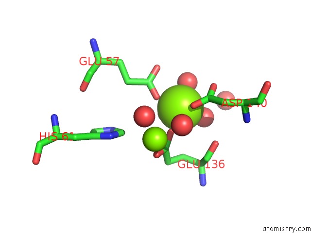
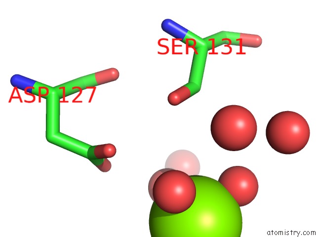
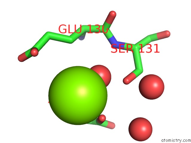
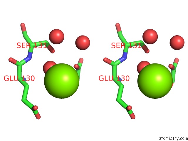
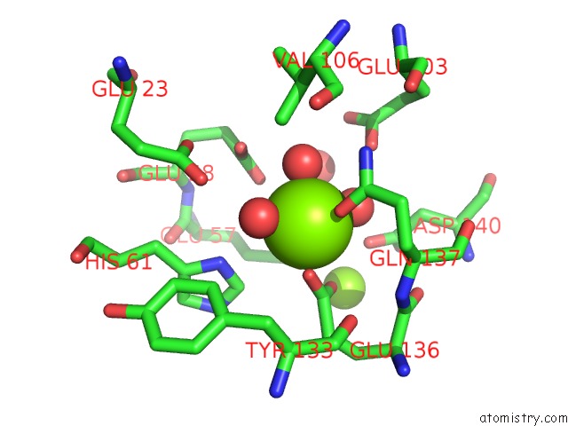
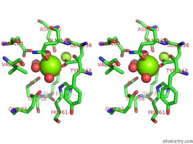
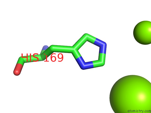
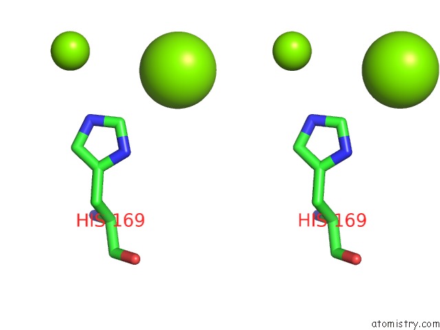
Magnesium binding site 1 out of 10 in 5xhm
Go back to
Magnesium binding site 1 out
of 10 in the Crystal Structure of Frog M-Ferritin D40A Mutant
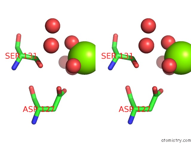
Mono view
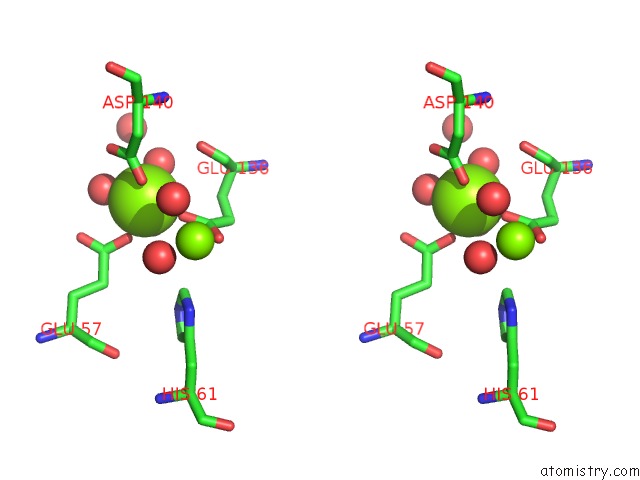
Stereo pair view
Mono view
Stereo pair view
A full contact list of Magnesium with other atoms in the Mg binding
site number 1 of Crystal Structure of Frog M-Ferritin D40A Mutant within 5.0Å range:
|
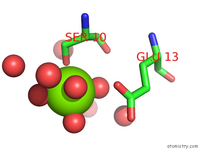
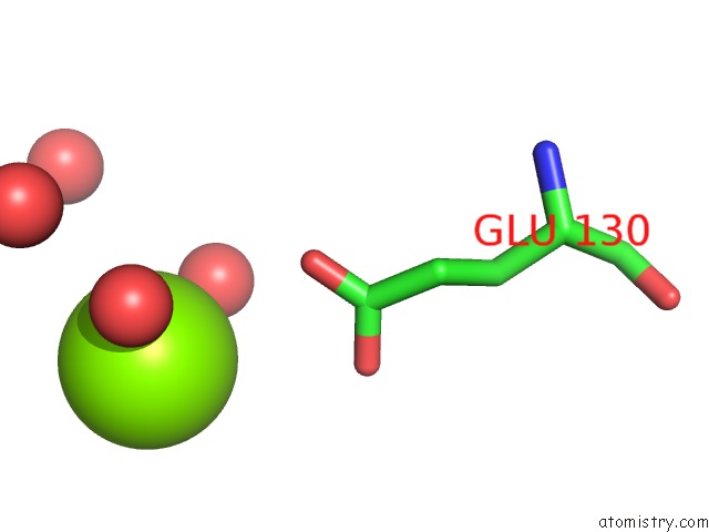
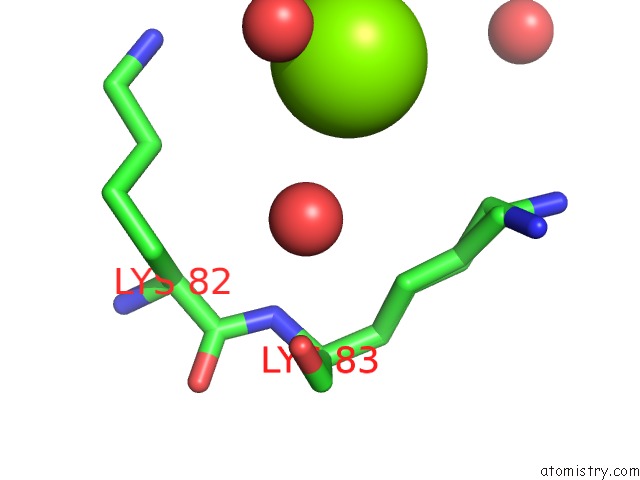
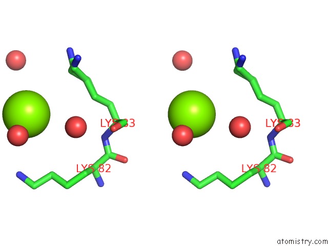
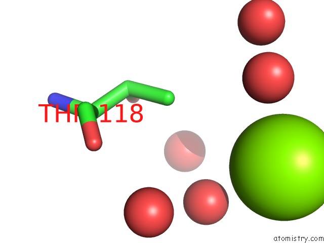
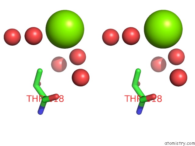
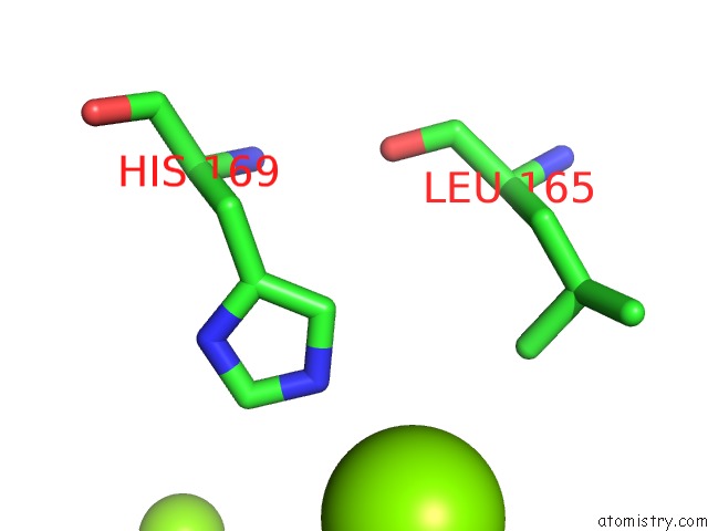
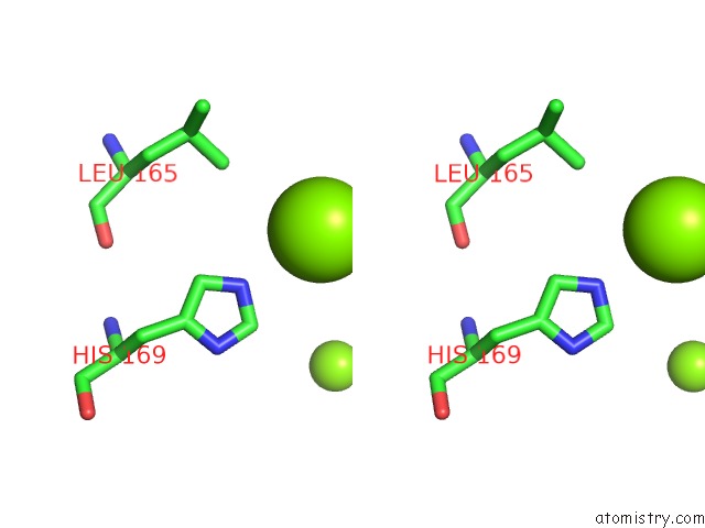
Magnesium binding site 2 out of 10 in 5xhm
Go back to
Magnesium binding site 2 out
of 10 in the Crystal Structure of Frog M-Ferritin D40A Mutant
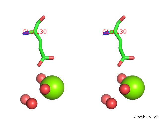
Mono view
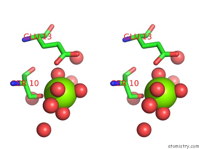
Stereo pair view
Mono view
Stereo pair view
A full contact list of Magnesium with other atoms in the Mg binding
site number 2 of Crystal Structure of Frog M-Ferritin D40A Mutant within 5.0Å range:
|
Magnesium binding site 3 out of 10 in 5xhm
Go back to
Magnesium binding site 3 out
of 10 in the Crystal Structure of Frog M-Ferritin D40A Mutant
Mono view
Stereo pair view
Mono view
Stereo pair view
A full contact list of Magnesium with other atoms in the Mg binding
site number 3 of Crystal Structure of Frog M-Ferritin D40A Mutant within 5.0Å range:
|
Magnesium binding site 4 out of 10 in 5xhm
Go back to
Magnesium binding site 4 out
of 10 in the Crystal Structure of Frog M-Ferritin D40A Mutant
Mono view
Stereo pair view
Mono view
Stereo pair view
A full contact list of Magnesium with other atoms in the Mg binding
site number 4 of Crystal Structure of Frog M-Ferritin D40A Mutant within 5.0Å range:
|
Magnesium binding site 5 out of 10 in 5xhm
Go back to
Magnesium binding site 5 out
of 10 in the Crystal Structure of Frog M-Ferritin D40A Mutant
Mono view
Stereo pair view
Mono view
Stereo pair view
A full contact list of Magnesium with other atoms in the Mg binding
site number 5 of Crystal Structure of Frog M-Ferritin D40A Mutant within 5.0Å range:
|
Magnesium binding site 6 out of 10 in 5xhm
Go back to
Magnesium binding site 6 out
of 10 in the Crystal Structure of Frog M-Ferritin D40A Mutant
Mono view
Stereo pair view
Mono view
Stereo pair view
A full contact list of Magnesium with other atoms in the Mg binding
site number 6 of Crystal Structure of Frog M-Ferritin D40A Mutant within 5.0Å range:
|
Magnesium binding site 7 out of 10 in 5xhm
Go back to
Magnesium binding site 7 out
of 10 in the Crystal Structure of Frog M-Ferritin D40A Mutant
Mono view
Stereo pair view
Mono view
Stereo pair view
A full contact list of Magnesium with other atoms in the Mg binding
site number 7 of Crystal Structure of Frog M-Ferritin D40A Mutant within 5.0Å range:
|
Magnesium binding site 8 out of 10 in 5xhm
Go back to
Magnesium binding site 8 out
of 10 in the Crystal Structure of Frog M-Ferritin D40A Mutant
Mono view
Stereo pair view
Mono view
Stereo pair view
A full contact list of Magnesium with other atoms in the Mg binding
site number 8 of Crystal Structure of Frog M-Ferritin D40A Mutant within 5.0Å range:
|
Magnesium binding site 9 out of 10 in 5xhm
Go back to
Magnesium binding site 9 out
of 10 in the Crystal Structure of Frog M-Ferritin D40A Mutant
Mono view
Stereo pair view
Mono view
Stereo pair view
A full contact list of Magnesium with other atoms in the Mg binding
site number 9 of Crystal Structure of Frog M-Ferritin D40A Mutant within 5.0Å range:
|
Magnesium binding site 10 out of 10 in 5xhm
Go back to
Magnesium binding site 10 out
of 10 in the Crystal Structure of Frog M-Ferritin D40A Mutant
Mono view
Stereo pair view
Mono view
Stereo pair view
A full contact list of Magnesium with other atoms in the Mg binding
site number 10 of Crystal Structure of Frog M-Ferritin D40A Mutant within 5.0Å range:
|
Reference:
B.Subhadarshanee,
A.Mohanty,
M.K.Jagdev,
D.Vasudevan,
R.K.Behera.
Surface Charge Dependent Separation of Modified and Hybrid Ferritin in Native Page: Impact of Lysine 104 Biochim. Biophys. Acta V.1865 1267 2017.
ISSN: ISSN 0006-3002
PubMed: 28739445
DOI: 10.1016/J.BBAPAP.2017.07.012
Page generated: Mon Sep 30 09:22:23 2024
ISSN: ISSN 0006-3002
PubMed: 28739445
DOI: 10.1016/J.BBAPAP.2017.07.012
Last articles
Zn in 9JYWZn in 9IR4
Zn in 9IR3
Zn in 9GMX
Zn in 9GMW
Zn in 9JEJ
Zn in 9ERF
Zn in 9ERE
Zn in 9EGV
Zn in 9EGW