Magnesium »
PDB 5xlf-5xuj »
5xm3 »
Magnesium in PDB 5xm3: Crystal Structure of Methanol Dehydrogenase From Methylophaga Aminisulfidivorans
Enzymatic activity of Crystal Structure of Methanol Dehydrogenase From Methylophaga Aminisulfidivorans
All present enzymatic activity of Crystal Structure of Methanol Dehydrogenase From Methylophaga Aminisulfidivorans:
1.1.2.7;
1.1.2.7;
Protein crystallography data
The structure of Crystal Structure of Methanol Dehydrogenase From Methylophaga Aminisulfidivorans, PDB code: 5xm3
was solved by
T.P.Cao,
J.M.Choi,
S.H.Lee,
with X-Ray Crystallography technique. A brief refinement statistics is given in the table below:
| Resolution Low / High (Å) | 34.72 / 1.70 |
| Space group | P 1 21 1 |
| Cell size a, b, c (Å), α, β, γ (°) | 63.926, 109.479, 95.554, 90.00, 100.48, 90.00 |
| R / Rfree (%) | 15.1 / 18.1 |
Magnesium Binding Sites:
The binding sites of Magnesium atom in the Crystal Structure of Methanol Dehydrogenase From Methylophaga Aminisulfidivorans
(pdb code 5xm3). This binding sites where shown within
5.0 Angstroms radius around Magnesium atom.
In total 2 binding sites of Magnesium where determined in the Crystal Structure of Methanol Dehydrogenase From Methylophaga Aminisulfidivorans, PDB code: 5xm3:
Jump to Magnesium binding site number: 1; 2;
In total 2 binding sites of Magnesium where determined in the Crystal Structure of Methanol Dehydrogenase From Methylophaga Aminisulfidivorans, PDB code: 5xm3:
Jump to Magnesium binding site number: 1; 2;
Magnesium binding site 1 out of 2 in 5xm3
Go back to
Magnesium binding site 1 out
of 2 in the Crystal Structure of Methanol Dehydrogenase From Methylophaga Aminisulfidivorans
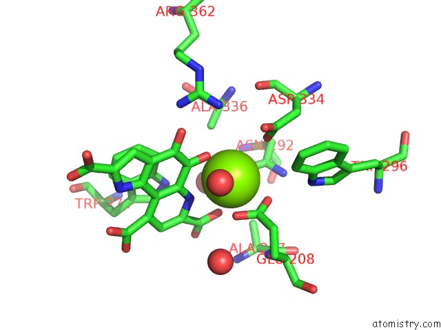
Mono view
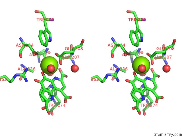
Stereo pair view
Mono view
Stereo pair view
A full contact list of Magnesium with other atoms in the Mg binding
site number 1 of Crystal Structure of Methanol Dehydrogenase From Methylophaga Aminisulfidivorans within 5.0Å range:
|
Magnesium binding site 2 out of 2 in 5xm3
Go back to
Magnesium binding site 2 out
of 2 in the Crystal Structure of Methanol Dehydrogenase From Methylophaga Aminisulfidivorans
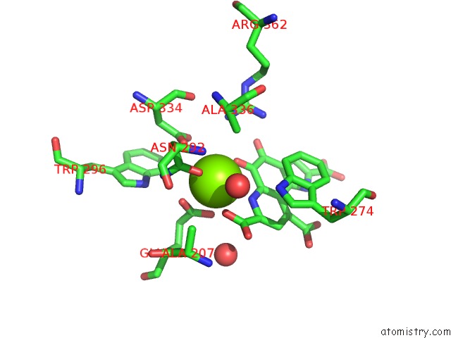
Mono view
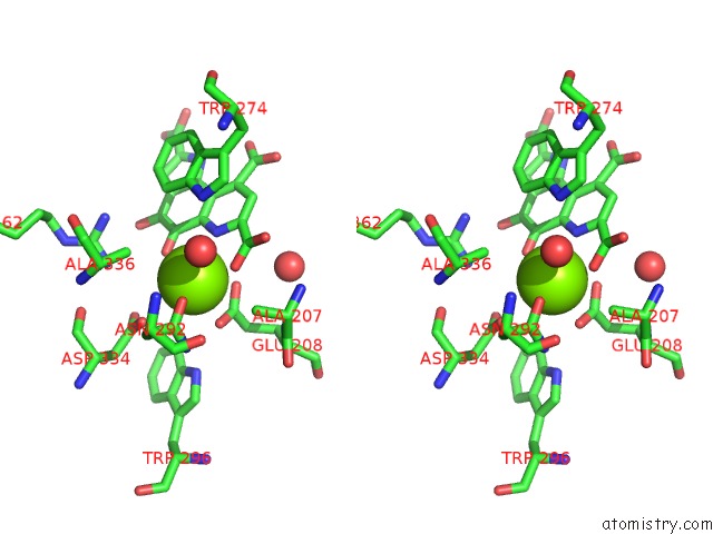
Stereo pair view
Mono view
Stereo pair view
A full contact list of Magnesium with other atoms in the Mg binding
site number 2 of Crystal Structure of Methanol Dehydrogenase From Methylophaga Aminisulfidivorans within 5.0Å range:
|
Reference:
T.P.Cao,
J.M.Choi,
S.W.Kim,
S.H.Lee.
The Crystal Structure of Methanol Dehydrogenase, A Quinoprotein From the Marine Methylotrophic Bacterium Methylophaga Aminisulfidivorans Mpt J. Microbiol. V. 56 246 2018.
ISSN: ESSN 1976-3794
PubMed: 29492864
DOI: 10.1007/S12275-018-7483-Y
Page generated: Mon Sep 30 09:34:20 2024
ISSN: ESSN 1976-3794
PubMed: 29492864
DOI: 10.1007/S12275-018-7483-Y
Last articles
Zn in 9J0NZn in 9J0O
Zn in 9J0P
Zn in 9FJX
Zn in 9EKB
Zn in 9C0F
Zn in 9CAH
Zn in 9CH0
Zn in 9CH3
Zn in 9CH1