Magnesium »
PDB 5xlg-5xus »
5xnx »
Magnesium in PDB 5xnx: Crystallographic Structure of the Enzymatically Active N-Terminal Domain of the Rel Protein From Mycobacterium Tuberculosis
Enzymatic activity of Crystallographic Structure of the Enzymatically Active N-Terminal Domain of the Rel Protein From Mycobacterium Tuberculosis
All present enzymatic activity of Crystallographic Structure of the Enzymatically Active N-Terminal Domain of the Rel Protein From Mycobacterium Tuberculosis:
2.7.6.5; 3.1.7.2;
2.7.6.5; 3.1.7.2;
Protein crystallography data
The structure of Crystallographic Structure of the Enzymatically Active N-Terminal Domain of the Rel Protein From Mycobacterium Tuberculosis, PDB code: 5xnx
was solved by
B.Singal,
A.M.Balakrishna,
M.S.S.Manimekalai,
W.Nartey,
G.Gruber,
with X-Ray Crystallography technique. A brief refinement statistics is given in the table below:
| Resolution Low / High (Å) | 10.02 / 3.70 |
| Space group | P 32 |
| Cell size a, b, c (Å), α, β, γ (°) | 161.710, 161.710, 75.560, 90.00, 90.00, 120.00 |
| R / Rfree (%) | 35 / 36.8 |
Magnesium Binding Sites:
The binding sites of Magnesium atom in the Crystallographic Structure of the Enzymatically Active N-Terminal Domain of the Rel Protein From Mycobacterium Tuberculosis
(pdb code 5xnx). This binding sites where shown within
5.0 Angstroms radius around Magnesium atom.
In total 3 binding sites of Magnesium where determined in the Crystallographic Structure of the Enzymatically Active N-Terminal Domain of the Rel Protein From Mycobacterium Tuberculosis, PDB code: 5xnx:
Jump to Magnesium binding site number: 1; 2; 3;
In total 3 binding sites of Magnesium where determined in the Crystallographic Structure of the Enzymatically Active N-Terminal Domain of the Rel Protein From Mycobacterium Tuberculosis, PDB code: 5xnx:
Jump to Magnesium binding site number: 1; 2; 3;
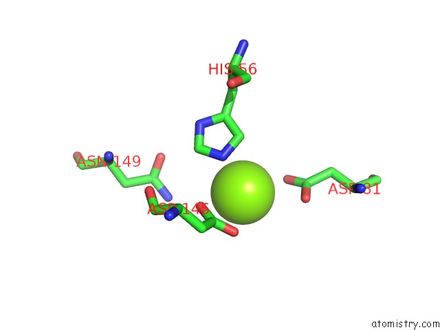
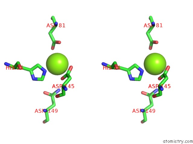
Magnesium binding site 1 out of 3 in 5xnx
Go back to
Magnesium binding site 1 out
of 3 in the Crystallographic Structure of the Enzymatically Active N-Terminal Domain of the Rel Protein From Mycobacterium Tuberculosis
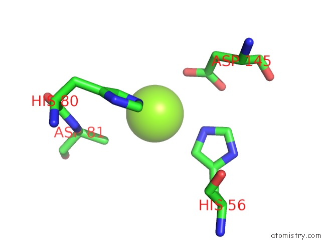
Mono view
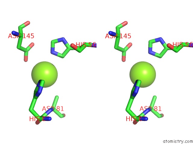
Stereo pair view
Mono view
Stereo pair view
A full contact list of Magnesium with other atoms in the Mg binding
site number 1 of Crystallographic Structure of the Enzymatically Active N-Terminal Domain of the Rel Protein From Mycobacterium Tuberculosis within 5.0Å range:
|
Magnesium binding site 2 out of 3 in 5xnx
Go back to
Magnesium binding site 2 out
of 3 in the Crystallographic Structure of the Enzymatically Active N-Terminal Domain of the Rel Protein From Mycobacterium Tuberculosis
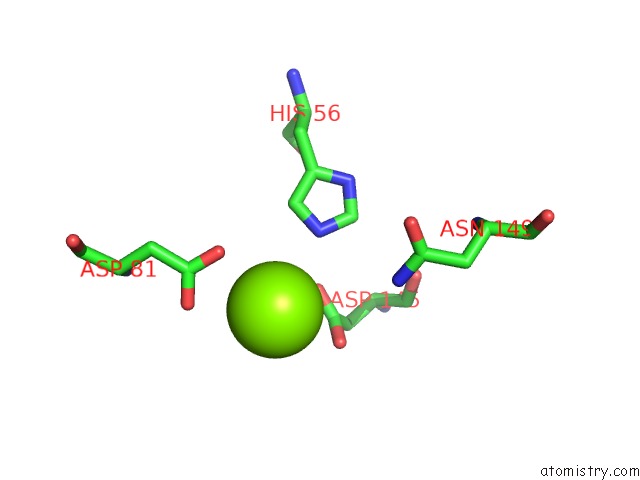
Mono view
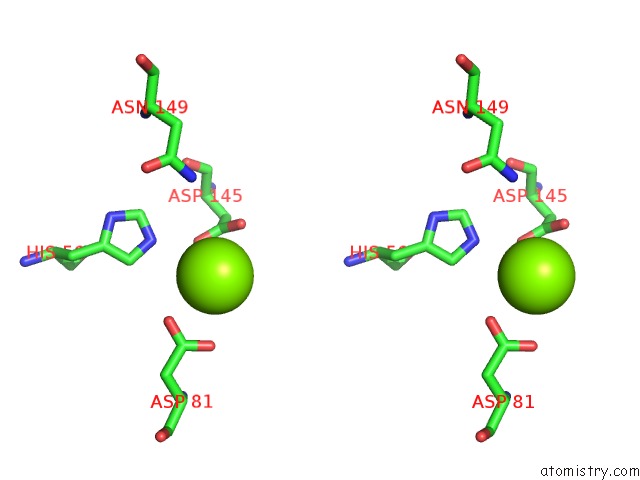
Stereo pair view
Mono view
Stereo pair view
A full contact list of Magnesium with other atoms in the Mg binding
site number 2 of Crystallographic Structure of the Enzymatically Active N-Terminal Domain of the Rel Protein From Mycobacterium Tuberculosis within 5.0Å range:
|
Magnesium binding site 3 out of 3 in 5xnx
Go back to
Magnesium binding site 3 out
of 3 in the Crystallographic Structure of the Enzymatically Active N-Terminal Domain of the Rel Protein From Mycobacterium Tuberculosis
Mono view
Stereo pair view
Mono view
Stereo pair view
A full contact list of Magnesium with other atoms in the Mg binding
site number 3 of Crystallographic Structure of the Enzymatically Active N-Terminal Domain of the Rel Protein From Mycobacterium Tuberculosis within 5.0Å range:
|
Reference:
B.Singal,
A.M.Balakrishna,
W.Nartey,
M.S.S.Manimekalai,
J.Jeyakanthan,
G.Gruber.
Crystallographic and Solution Structure of the N-Terminal Domain of the Rel Protein From Mycobacterium Tuberculosis Febs Lett. V. 591 2323 2017.
ISSN: ISSN 1873-3468
PubMed: 28672070
DOI: 10.1002/1873-3468.12739
Page generated: Mon Sep 30 09:36:03 2024
ISSN: ISSN 1873-3468
PubMed: 28672070
DOI: 10.1002/1873-3468.12739
Last articles
Zn in 9MJ5Zn in 9HNW
Zn in 9G0L
Zn in 9FNE
Zn in 9DZN
Zn in 9E0I
Zn in 9D32
Zn in 9DAK
Zn in 8ZXC
Zn in 8ZUF