Magnesium »
PDB 5xlf-5xuj »
5xp0 »
Magnesium in PDB 5xp0: Crystal Structure of Master Biofilm Regulator Csgd Regulatory Domain
Protein crystallography data
The structure of Crystal Structure of Master Biofilm Regulator Csgd Regulatory Domain, PDB code: 5xp0
was solved by
Y.Wen,
Z.Ouyang,
with X-Ray Crystallography technique. A brief refinement statistics is given in the table below:
| Resolution Low / High (Å) | 44.36 / 2.00 |
| Space group | P 41 21 2 |
| Cell size a, b, c (Å), α, β, γ (°) | 57.758, 57.758, 207.742, 90.00, 90.00, 90.00 |
| R / Rfree (%) | 20.6 / 23.8 |
Magnesium Binding Sites:
The binding sites of Magnesium atom in the Crystal Structure of Master Biofilm Regulator Csgd Regulatory Domain
(pdb code 5xp0). This binding sites where shown within
5.0 Angstroms radius around Magnesium atom.
In total 2 binding sites of Magnesium where determined in the Crystal Structure of Master Biofilm Regulator Csgd Regulatory Domain, PDB code: 5xp0:
Jump to Magnesium binding site number: 1; 2;
In total 2 binding sites of Magnesium where determined in the Crystal Structure of Master Biofilm Regulator Csgd Regulatory Domain, PDB code: 5xp0:
Jump to Magnesium binding site number: 1; 2;
Magnesium binding site 1 out of 2 in 5xp0
Go back to
Magnesium binding site 1 out
of 2 in the Crystal Structure of Master Biofilm Regulator Csgd Regulatory Domain
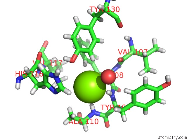
Mono view
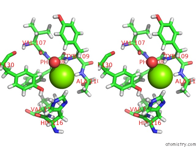
Stereo pair view
Mono view
Stereo pair view
A full contact list of Magnesium with other atoms in the Mg binding
site number 1 of Crystal Structure of Master Biofilm Regulator Csgd Regulatory Domain within 5.0Å range:
|
Magnesium binding site 2 out of 2 in 5xp0
Go back to
Magnesium binding site 2 out
of 2 in the Crystal Structure of Master Biofilm Regulator Csgd Regulatory Domain
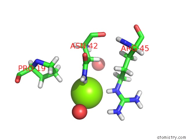
Mono view
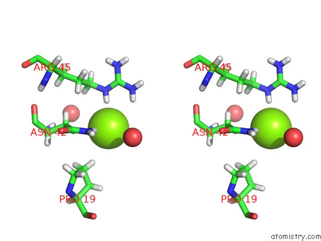
Stereo pair view
Mono view
Stereo pair view
A full contact list of Magnesium with other atoms in the Mg binding
site number 2 of Crystal Structure of Master Biofilm Regulator Csgd Regulatory Domain within 5.0Å range:
|
Reference:
Y.Wen,
Z.Ouyang,
B.Devreese,
W.He,
Y.Shao,
W.Lu,
F.Zheng.
Crystal Structure of Master Biofilm Regulator Csgd Regulatory Domain Reveals An Atypical Receiver Domain. Protein Sci. V. 26 2073 2017.
ISSN: ESSN 1469-896X
PubMed: 28758290
DOI: 10.1002/PRO.3245
Page generated: Mon Sep 30 09:37:23 2024
ISSN: ESSN 1469-896X
PubMed: 28758290
DOI: 10.1002/PRO.3245
Last articles
Zn in 9J0NZn in 9J0O
Zn in 9J0P
Zn in 9FJX
Zn in 9EKB
Zn in 9C0F
Zn in 9CAH
Zn in 9CH0
Zn in 9CH3
Zn in 9CH1