Magnesium »
PDB 5xlg-5xus »
5xuj »
Magnesium in PDB 5xuj: Crystal Structure of PDE10A in Complex with 7-(4-Chlorophenyl)-2- Methylpyrazolo[1,5-A]Pyrimidine
Enzymatic activity of Crystal Structure of PDE10A in Complex with 7-(4-Chlorophenyl)-2- Methylpyrazolo[1,5-A]Pyrimidine
All present enzymatic activity of Crystal Structure of PDE10A in Complex with 7-(4-Chlorophenyl)-2- Methylpyrazolo[1,5-A]Pyrimidine:
3.1.4.17; 3.1.4.35;
3.1.4.17; 3.1.4.35;
Protein crystallography data
The structure of Crystal Structure of PDE10A in Complex with 7-(4-Chlorophenyl)-2- Methylpyrazolo[1,5-A]Pyrimidine, PDB code: 5xuj
was solved by
Y.Amano,
K.Honbou,
with X-Ray Crystallography technique. A brief refinement statistics is given in the table below:
| Resolution Low / High (Å) | 78.80 / 2.44 |
| Space group | P 21 21 21 |
| Cell size a, b, c (Å), α, β, γ (°) | 50.339, 81.299, 157.597, 90.00, 90.00, 90.00 |
| R / Rfree (%) | 19.8 / 28 |
Other elements in 5xuj:
The structure of Crystal Structure of PDE10A in Complex with 7-(4-Chlorophenyl)-2- Methylpyrazolo[1,5-A]Pyrimidine also contains other interesting chemical elements:
| Chlorine | (Cl) | 1 atom |
| Zinc | (Zn) | 2 atoms |
Magnesium Binding Sites:
The binding sites of Magnesium atom in the Crystal Structure of PDE10A in Complex with 7-(4-Chlorophenyl)-2- Methylpyrazolo[1,5-A]Pyrimidine
(pdb code 5xuj). This binding sites where shown within
5.0 Angstroms radius around Magnesium atom.
In total 2 binding sites of Magnesium where determined in the Crystal Structure of PDE10A in Complex with 7-(4-Chlorophenyl)-2- Methylpyrazolo[1,5-A]Pyrimidine, PDB code: 5xuj:
Jump to Magnesium binding site number: 1; 2;
In total 2 binding sites of Magnesium where determined in the Crystal Structure of PDE10A in Complex with 7-(4-Chlorophenyl)-2- Methylpyrazolo[1,5-A]Pyrimidine, PDB code: 5xuj:
Jump to Magnesium binding site number: 1; 2;
Magnesium binding site 1 out of 2 in 5xuj
Go back to
Magnesium binding site 1 out
of 2 in the Crystal Structure of PDE10A in Complex with 7-(4-Chlorophenyl)-2- Methylpyrazolo[1,5-A]Pyrimidine
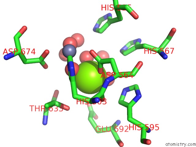
Mono view
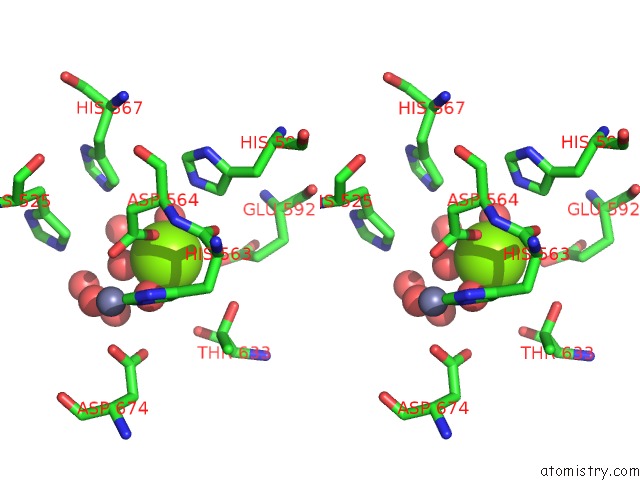
Stereo pair view
Mono view
Stereo pair view
A full contact list of Magnesium with other atoms in the Mg binding
site number 1 of Crystal Structure of PDE10A in Complex with 7-(4-Chlorophenyl)-2- Methylpyrazolo[1,5-A]Pyrimidine within 5.0Å range:
|
Magnesium binding site 2 out of 2 in 5xuj
Go back to
Magnesium binding site 2 out
of 2 in the Crystal Structure of PDE10A in Complex with 7-(4-Chlorophenyl)-2- Methylpyrazolo[1,5-A]Pyrimidine
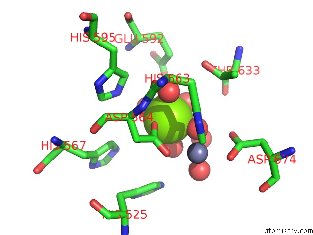
Mono view
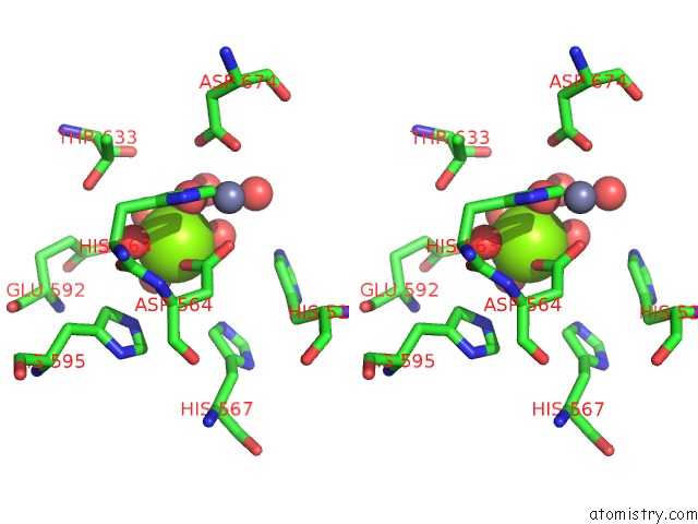
Stereo pair view
Mono view
Stereo pair view
A full contact list of Magnesium with other atoms in the Mg binding
site number 2 of Crystal Structure of PDE10A in Complex with 7-(4-Chlorophenyl)-2- Methylpyrazolo[1,5-A]Pyrimidine within 5.0Å range:
|
Reference:
A.Chino,
R.Seo,
Y.Amano,
I.Namatame,
W.Hamaguchi,
K.Honbou,
T.Mihara,
M.Yamazaki,
M.Tomishima,
N.Masuda.
Fragment-Based Discovery of Pyrimido[1,2-B]Indazole PDE10A Inhibitors. Chem. Pharm. Bull. V. 66 286 2018.
ISSN: ISSN 1347-5223
PubMed: 29491261
DOI: 10.1248/CPB.C17-00836
Page generated: Mon Sep 30 09:44:23 2024
ISSN: ISSN 1347-5223
PubMed: 29491261
DOI: 10.1248/CPB.C17-00836
Last articles
F in 4JU4F in 4JTZ
F in 4JTY
F in 4JTW
F in 4JSX
F in 4JSV
F in 4JTQ
F in 4JSC
F in 4JSJ
F in 4JSM