Magnesium »
PDB 5xus-5y4i »
5xxl »
Magnesium in PDB 5xxl: Crystal Structure of GH3 Beta-Glucosidase From Bacteroides Thetaiotaomicron
Protein crystallography data
The structure of Crystal Structure of GH3 Beta-Glucosidase From Bacteroides Thetaiotaomicron, PDB code: 5xxl
was solved by
M.Nakajima,
R.Ishiguro,
N.Tanaka,
K.Abe,
T.Maeda,
A.Miyanaga,
Y.Takahash,
N.Sugimoto,
H.Nakai,
H.Taguchi,
with X-Ray Crystallography technique. A brief refinement statistics is given in the table below:
| Resolution Low / High (Å) | 29.94 / 1.60 |
| Space group | C 2 2 21 |
| Cell size a, b, c (Å), α, β, γ (°) | 81.940, 167.880, 224.090, 90.00, 90.00, 90.00 |
| R / Rfree (%) | 20.4 / 22.7 |
Magnesium Binding Sites:
The binding sites of Magnesium atom in the Crystal Structure of GH3 Beta-Glucosidase From Bacteroides Thetaiotaomicron
(pdb code 5xxl). This binding sites where shown within
5.0 Angstroms radius around Magnesium atom.
In total 2 binding sites of Magnesium where determined in the Crystal Structure of GH3 Beta-Glucosidase From Bacteroides Thetaiotaomicron, PDB code: 5xxl:
Jump to Magnesium binding site number: 1; 2;
In total 2 binding sites of Magnesium where determined in the Crystal Structure of GH3 Beta-Glucosidase From Bacteroides Thetaiotaomicron, PDB code: 5xxl:
Jump to Magnesium binding site number: 1; 2;
Magnesium binding site 1 out of 2 in 5xxl
Go back to
Magnesium binding site 1 out
of 2 in the Crystal Structure of GH3 Beta-Glucosidase From Bacteroides Thetaiotaomicron
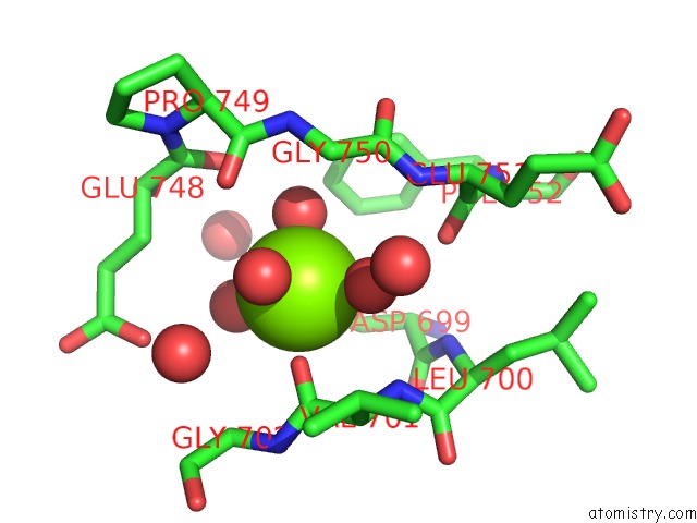
Mono view
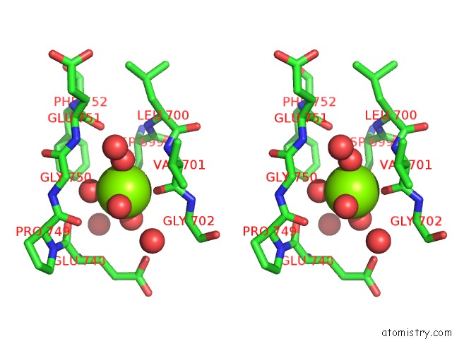
Stereo pair view
Mono view
Stereo pair view
A full contact list of Magnesium with other atoms in the Mg binding
site number 1 of Crystal Structure of GH3 Beta-Glucosidase From Bacteroides Thetaiotaomicron within 5.0Å range:
|
Magnesium binding site 2 out of 2 in 5xxl
Go back to
Magnesium binding site 2 out
of 2 in the Crystal Structure of GH3 Beta-Glucosidase From Bacteroides Thetaiotaomicron
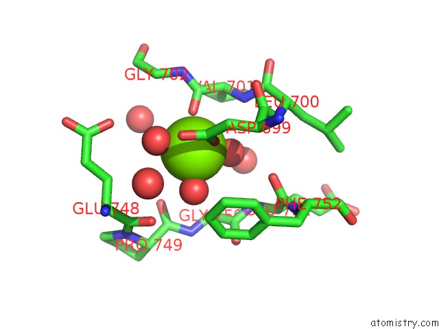
Mono view
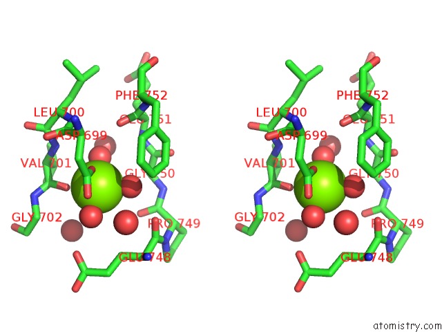
Stereo pair view
Mono view
Stereo pair view
A full contact list of Magnesium with other atoms in the Mg binding
site number 2 of Crystal Structure of GH3 Beta-Glucosidase From Bacteroides Thetaiotaomicron within 5.0Å range:
|
Reference:
R.Ishiguro,
N.Tanaka,
K.Abe,
M.Nakajima,
T.Maeda,
A.Miyanaga,
Y.Takahashi,
N.Sugimoto,
H.Nakai,
H.Taguchi.
Function and Structure Relationships of A Beta-1,2-Glucooligosaccharide-Degrading Beta-Glucosidase. Febs Lett. V. 591 3926 2017.
ISSN: ISSN 1873-3468
PubMed: 29131329
DOI: 10.1002/1873-3468.12911
Page generated: Mon Sep 30 10:37:54 2024
ISSN: ISSN 1873-3468
PubMed: 29131329
DOI: 10.1002/1873-3468.12911
Last articles
Zn in 9J0NZn in 9J0O
Zn in 9J0P
Zn in 9FJX
Zn in 9EKB
Zn in 9C0F
Zn in 9CAH
Zn in 9CH0
Zn in 9CH3
Zn in 9CH1