Magnesium »
PDB 5ylu-5yve »
5yth »
Magnesium in PDB 5yth: Structure of Large Fragment of Dna Polymerase I From Thermus Aquaticus Host-Guest Complex with the Unnatural Base M-Fc Pair with Dg
Enzymatic activity of Structure of Large Fragment of Dna Polymerase I From Thermus Aquaticus Host-Guest Complex with the Unnatural Base M-Fc Pair with Dg
All present enzymatic activity of Structure of Large Fragment of Dna Polymerase I From Thermus Aquaticus Host-Guest Complex with the Unnatural Base M-Fc Pair with Dg:
2.7.7.7;
2.7.7.7;
Protein crystallography data
The structure of Structure of Large Fragment of Dna Polymerase I From Thermus Aquaticus Host-Guest Complex with the Unnatural Base M-Fc Pair with Dg, PDB code: 5yth
was solved by
H.Zeng,
M.Mondal,
R.Y.Song,
J.Zhang,
B.Xia,
Y.Q.Gao,
C.Q.Yi,
with X-Ray Crystallography technique. A brief refinement statistics is given in the table below:
| Resolution Low / High (Å) | 94.63 / 2.53 |
| Space group | P 31 2 1 |
| Cell size a, b, c (Å), α, β, γ (°) | 109.273, 109.273, 91.119, 90.00, 90.00, 120.00 |
| R / Rfree (%) | 20.4 / 27.5 |
Magnesium Binding Sites:
The binding sites of Magnesium atom in the Structure of Large Fragment of Dna Polymerase I From Thermus Aquaticus Host-Guest Complex with the Unnatural Base M-Fc Pair with Dg
(pdb code 5yth). This binding sites where shown within
5.0 Angstroms radius around Magnesium atom.
In total 2 binding sites of Magnesium where determined in the Structure of Large Fragment of Dna Polymerase I From Thermus Aquaticus Host-Guest Complex with the Unnatural Base M-Fc Pair with Dg, PDB code: 5yth:
Jump to Magnesium binding site number: 1; 2;
In total 2 binding sites of Magnesium where determined in the Structure of Large Fragment of Dna Polymerase I From Thermus Aquaticus Host-Guest Complex with the Unnatural Base M-Fc Pair with Dg, PDB code: 5yth:
Jump to Magnesium binding site number: 1; 2;
Magnesium binding site 1 out of 2 in 5yth
Go back to
Magnesium binding site 1 out
of 2 in the Structure of Large Fragment of Dna Polymerase I From Thermus Aquaticus Host-Guest Complex with the Unnatural Base M-Fc Pair with Dg
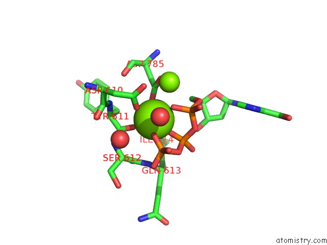
Mono view
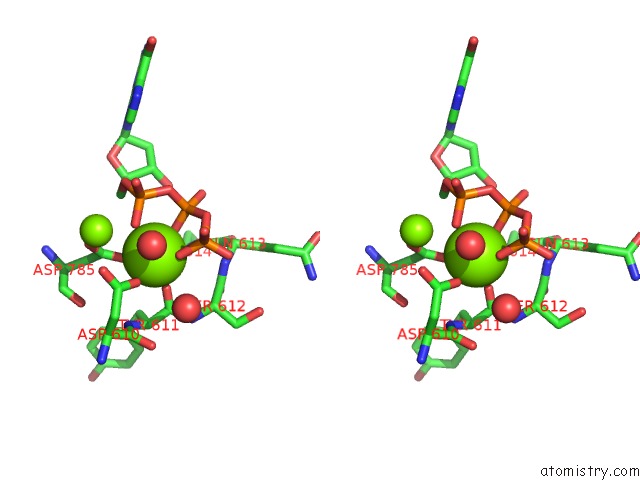
Stereo pair view
Mono view
Stereo pair view
A full contact list of Magnesium with other atoms in the Mg binding
site number 1 of Structure of Large Fragment of Dna Polymerase I From Thermus Aquaticus Host-Guest Complex with the Unnatural Base M-Fc Pair with Dg within 5.0Å range:
|
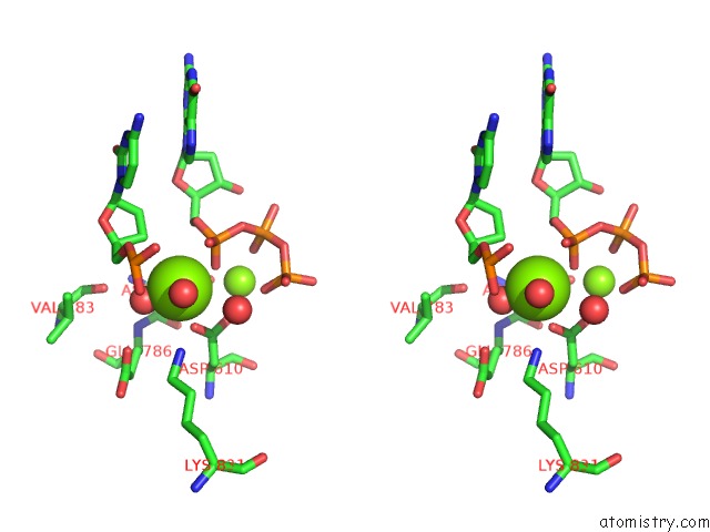
Magnesium binding site 2 out of 2 in 5yth
Go back to
Magnesium binding site 2 out
of 2 in the Structure of Large Fragment of Dna Polymerase I From Thermus Aquaticus Host-Guest Complex with the Unnatural Base M-Fc Pair with Dg
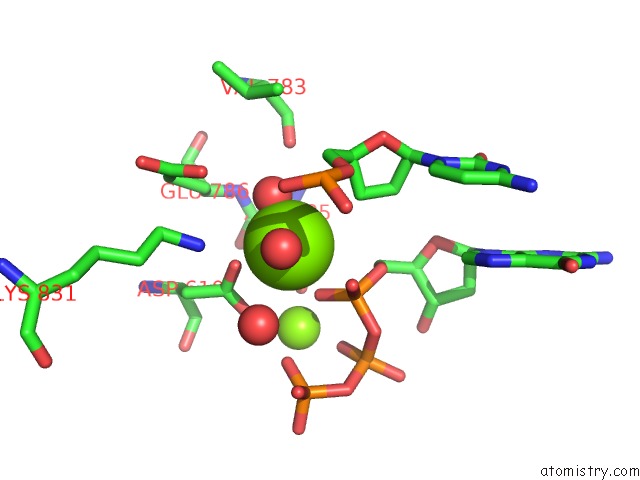
Mono view
Stereo pair view
Mono view
Stereo pair view
A full contact list of Magnesium with other atoms in the Mg binding
site number 2 of Structure of Large Fragment of Dna Polymerase I From Thermus Aquaticus Host-Guest Complex with the Unnatural Base M-Fc Pair with Dg within 5.0Å range:
|
Reference:
H.Zeng,
M.Mondal,
R.Song,
J.Zhang,
B.Xia,
M.Liu,
C.Zhu,
B.He,
Y.Q.Gao,
C.Yi.
Unnatural Cytosine Bases Recognized As Thymines By Dna Polymerases By the Formation of the Watson-Crick Geometry. Angew. Chem. Int. Ed. Engl. V. 58 130 2019.
ISSN: ESSN 1521-3773
PubMed: 30407705
DOI: 10.1002/ANIE.201807845
Page generated: Mon Sep 30 11:28:18 2024
ISSN: ESSN 1521-3773
PubMed: 30407705
DOI: 10.1002/ANIE.201807845
Last articles
Zn in 9J0NZn in 9J0O
Zn in 9J0P
Zn in 9FJX
Zn in 9EKB
Zn in 9C0F
Zn in 9CAH
Zn in 9CH0
Zn in 9CH3
Zn in 9CH1