Magnesium »
PDB 5yw2-5z68 »
5yws »
Magnesium in PDB 5yws: Crystal Structure of TREX1 in Complex with A Y Structured Dna
Enzymatic activity of Crystal Structure of TREX1 in Complex with A Y Structured Dna
All present enzymatic activity of Crystal Structure of TREX1 in Complex with A Y Structured Dna:
3.1.11.2;
3.1.11.2;
Protein crystallography data
The structure of Crystal Structure of TREX1 in Complex with A Y Structured Dna, PDB code: 5yws
was solved by
Y.Y.Hsiao,
with X-Ray Crystallography technique. A brief refinement statistics is given in the table below:
| Resolution Low / High (Å) | 29.33 / 2.00 |
| Space group | P 21 21 21 |
| Cell size a, b, c (Å), α, β, γ (°) | 59.532, 96.604, 101.112, 90.00, 90.00, 90.00 |
| R / Rfree (%) | 18.9 / 22.6 |
Magnesium Binding Sites:
The binding sites of Magnesium atom in the Crystal Structure of TREX1 in Complex with A Y Structured Dna
(pdb code 5yws). This binding sites where shown within
5.0 Angstroms radius around Magnesium atom.
In total 4 binding sites of Magnesium where determined in the Crystal Structure of TREX1 in Complex with A Y Structured Dna, PDB code: 5yws:
Jump to Magnesium binding site number: 1; 2; 3; 4;
In total 4 binding sites of Magnesium where determined in the Crystal Structure of TREX1 in Complex with A Y Structured Dna, PDB code: 5yws:
Jump to Magnesium binding site number: 1; 2; 3; 4;
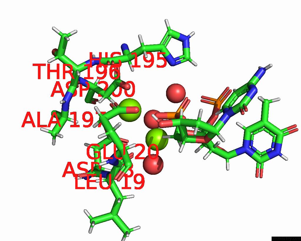
Magnesium binding site 1 out of 4 in 5yws
Go back to
Magnesium binding site 1 out
of 4 in the Crystal Structure of TREX1 in Complex with A Y Structured Dna
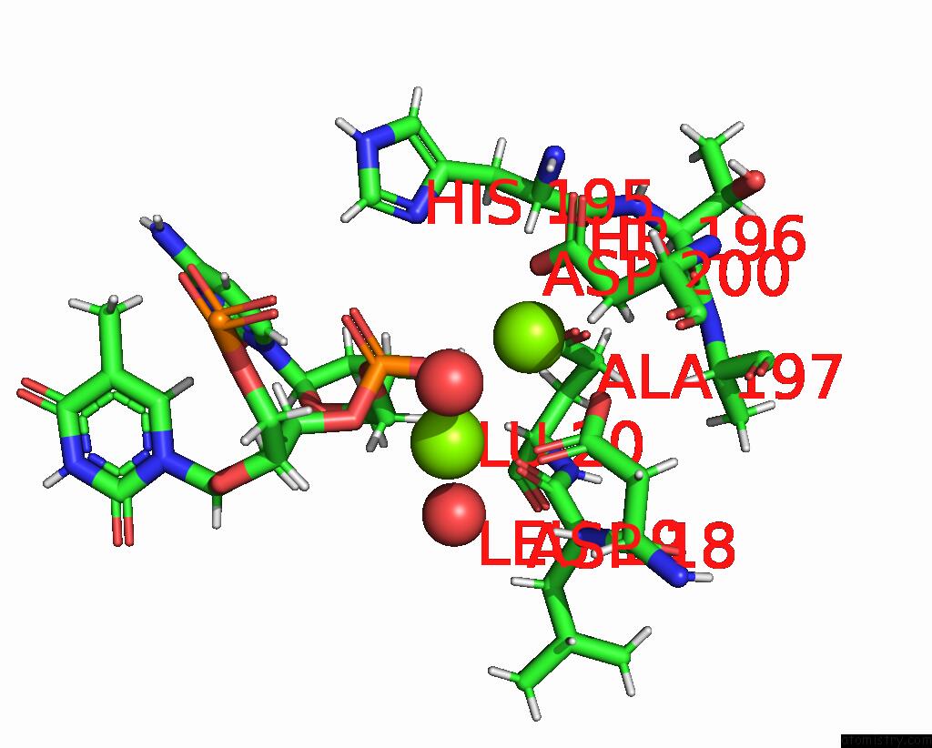
Mono view
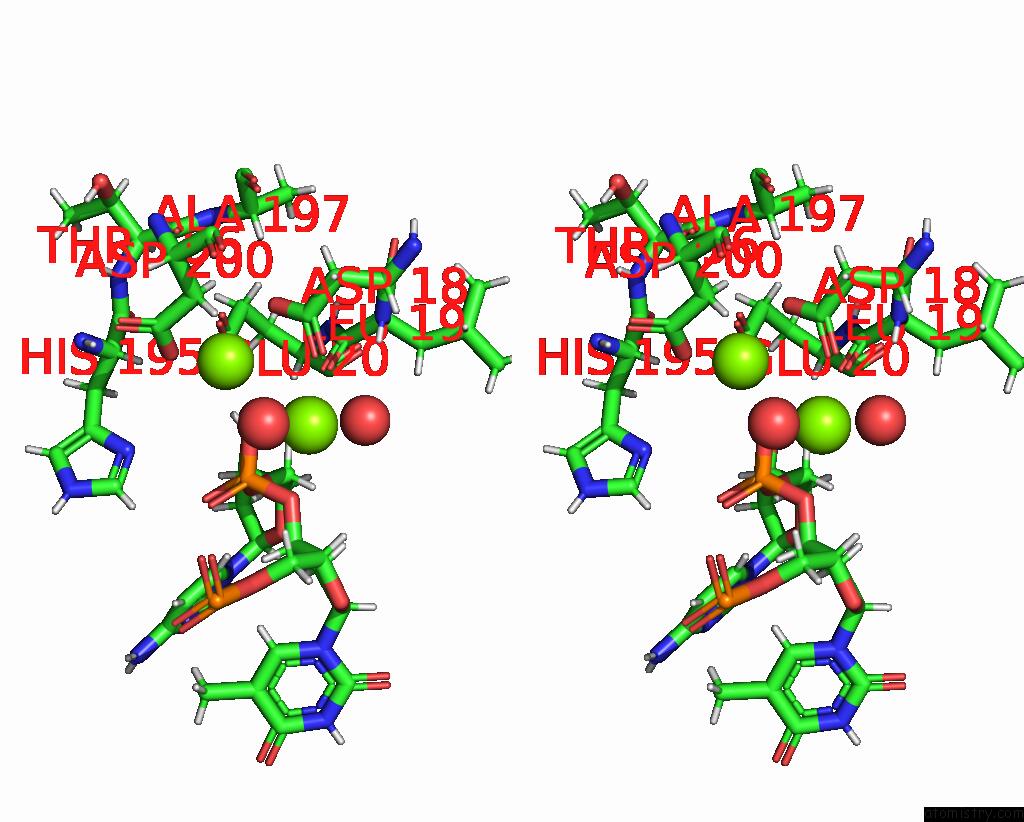
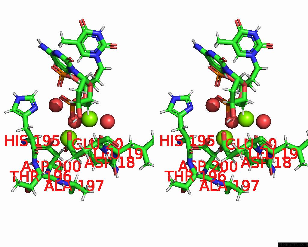
Stereo pair view
Mono view
Stereo pair view
A full contact list of Magnesium with other atoms in the Mg binding
site number 1 of Crystal Structure of TREX1 in Complex with A Y Structured Dna within 5.0Å range:
|
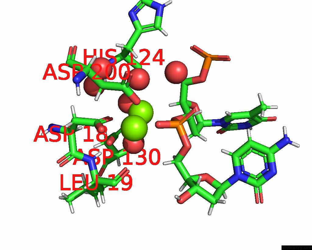
Magnesium binding site 2 out of 4 in 5yws
Go back to
Magnesium binding site 2 out
of 4 in the Crystal Structure of TREX1 in Complex with A Y Structured Dna
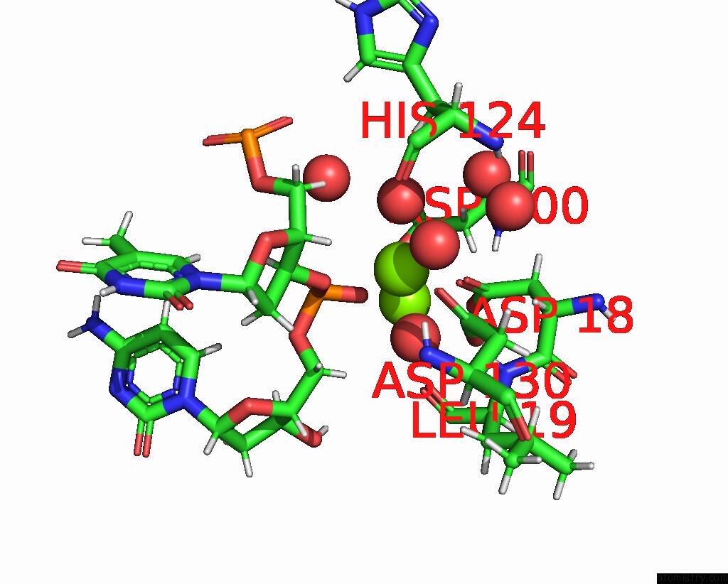
Mono view
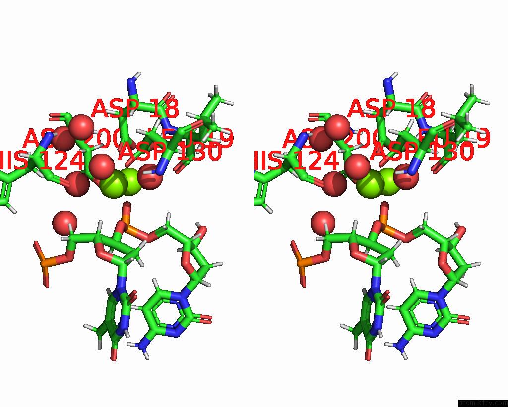
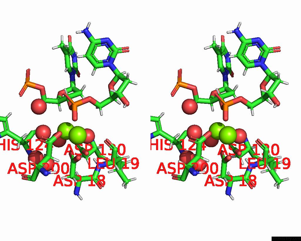
Stereo pair view
Mono view
Stereo pair view
A full contact list of Magnesium with other atoms in the Mg binding
site number 2 of Crystal Structure of TREX1 in Complex with A Y Structured Dna within 5.0Å range:
|
Magnesium binding site 3 out of 4 in 5yws
Go back to
Magnesium binding site 3 out
of 4 in the Crystal Structure of TREX1 in Complex with A Y Structured Dna
Mono view
Stereo pair view
Mono view
Stereo pair view
A full contact list of Magnesium with other atoms in the Mg binding
site number 3 of Crystal Structure of TREX1 in Complex with A Y Structured Dna within 5.0Å range:
|
Magnesium binding site 4 out of 4 in 5yws
Go back to
Magnesium binding site 4 out
of 4 in the Crystal Structure of TREX1 in Complex with A Y Structured Dna
Mono view
Stereo pair view
Mono view
Stereo pair view
A full contact list of Magnesium with other atoms in the Mg binding
site number 4 of Crystal Structure of TREX1 in Complex with A Y Structured Dna within 5.0Å range:
|
Reference:
K.W.Huang,
T.C.Liu,
R.Y.Liang,
L.Y.Chu,
H.L.Cheng,
J.W.Chu,
Y.Y.Hsiao.
Structural Basis For Overhang Excision and Terminal Unwinding of Dna Duplexes By TREX1 Plos Biol. V. 16 05653 2018.
ISSN: ESSN 1545-7885
PubMed: 29734329
DOI: 10.1371/JOURNAL.PBIO.2005653
Page generated: Mon Sep 30 11:37:02 2024
ISSN: ESSN 1545-7885
PubMed: 29734329
DOI: 10.1371/JOURNAL.PBIO.2005653
Last articles
Zn in 9MJ5Zn in 9HNW
Zn in 9G0L
Zn in 9FNE
Zn in 9DZN
Zn in 9E0I
Zn in 9D32
Zn in 9DAK
Zn in 8ZXC
Zn in 8ZUF