Magnesium »
PDB 5yw2-5z68 »
5z2w »
Magnesium in PDB 5z2w: Crystal Structure of the Bacterial Cell Division Protein Ftsq and Ftsb
Protein crystallography data
The structure of Crystal Structure of the Bacterial Cell Division Protein Ftsq and Ftsb, PDB code: 5z2w
was solved by
Y.Choi,
H.J.Yoon,
H.H.Lee,
with X-Ray Crystallography technique. A brief refinement statistics is given in the table below:
| Resolution Low / High (Å) | 36.47 / 3.00 |
| Space group | P 1 2 1 |
| Cell size a, b, c (Å), α, β, γ (°) | 63.557, 39.306, 73.191, 90.00, 94.77, 90.00 |
| R / Rfree (%) | 20.7 / 27.5 |
Magnesium Binding Sites:
The binding sites of Magnesium atom in the Crystal Structure of the Bacterial Cell Division Protein Ftsq and Ftsb
(pdb code 5z2w). This binding sites where shown within
5.0 Angstroms radius around Magnesium atom.
In total 2 binding sites of Magnesium where determined in the Crystal Structure of the Bacterial Cell Division Protein Ftsq and Ftsb, PDB code: 5z2w:
Jump to Magnesium binding site number: 1; 2;
In total 2 binding sites of Magnesium where determined in the Crystal Structure of the Bacterial Cell Division Protein Ftsq and Ftsb, PDB code: 5z2w:
Jump to Magnesium binding site number: 1; 2;
Magnesium binding site 1 out of 2 in 5z2w
Go back to
Magnesium binding site 1 out
of 2 in the Crystal Structure of the Bacterial Cell Division Protein Ftsq and Ftsb
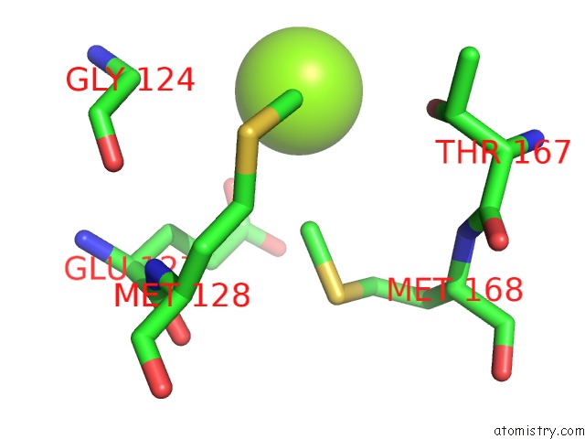
Mono view
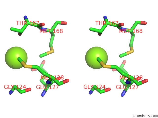
Stereo pair view
Mono view
Stereo pair view
A full contact list of Magnesium with other atoms in the Mg binding
site number 1 of Crystal Structure of the Bacterial Cell Division Protein Ftsq and Ftsb within 5.0Å range:
|
Magnesium binding site 2 out of 2 in 5z2w
Go back to
Magnesium binding site 2 out
of 2 in the Crystal Structure of the Bacterial Cell Division Protein Ftsq and Ftsb
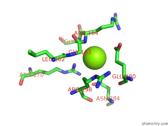
Mono view
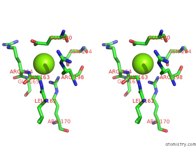
Stereo pair view
Mono view
Stereo pair view
A full contact list of Magnesium with other atoms in the Mg binding
site number 2 of Crystal Structure of the Bacterial Cell Division Protein Ftsq and Ftsb within 5.0Å range:
|
Reference:
Y.Choi,
J.Kim,
H.J.Yoon,
K.S.Jin,
S.Ryu,
H.H.Lee.
Structural Insights Into the Ftsq/Ftsb/Ftsl Complex, A Key Component of the Divisome. Sci Rep V. 8 18061 2018.
ISSN: ESSN 2045-2322
PubMed: 30584256
DOI: 10.1038/S41598-018-36001-2
Page generated: Mon Sep 30 11:41:29 2024
ISSN: ESSN 2045-2322
PubMed: 30584256
DOI: 10.1038/S41598-018-36001-2
Last articles
Zn in 9MJ5Zn in 9HNW
Zn in 9G0L
Zn in 9FNE
Zn in 9DZN
Zn in 9E0I
Zn in 9D32
Zn in 9DAK
Zn in 8ZXC
Zn in 8ZUF