Magnesium »
PDB 6a1m-6aad »
6a20 »
Magnesium in PDB 6a20: Crystal Structure of Auto-Inhibited Kinesin-3 KIF13B
Protein crystallography data
The structure of Crystal Structure of Auto-Inhibited Kinesin-3 KIF13B, PDB code: 6a20
was solved by
J.Q.Ren,
S.Wang,
W.Feng,
with X-Ray Crystallography technique. A brief refinement statistics is given in the table below:
| Resolution Low / High (Å) | 30.64 / 2.40 |
| Space group | P 32 |
| Cell size a, b, c (Å), α, β, γ (°) | 75.070, 75.070, 91.710, 90.00, 90.00, 120.00 |
| R / Rfree (%) | 19.3 / 24.9 |
Magnesium Binding Sites:
The binding sites of Magnesium atom in the Crystal Structure of Auto-Inhibited Kinesin-3 KIF13B
(pdb code 6a20). This binding sites where shown within
5.0 Angstroms radius around Magnesium atom.
In total 2 binding sites of Magnesium where determined in the Crystal Structure of Auto-Inhibited Kinesin-3 KIF13B, PDB code: 6a20:
Jump to Magnesium binding site number: 1; 2;
In total 2 binding sites of Magnesium where determined in the Crystal Structure of Auto-Inhibited Kinesin-3 KIF13B, PDB code: 6a20:
Jump to Magnesium binding site number: 1; 2;
Magnesium binding site 1 out of 2 in 6a20
Go back to
Magnesium binding site 1 out
of 2 in the Crystal Structure of Auto-Inhibited Kinesin-3 KIF13B

Mono view
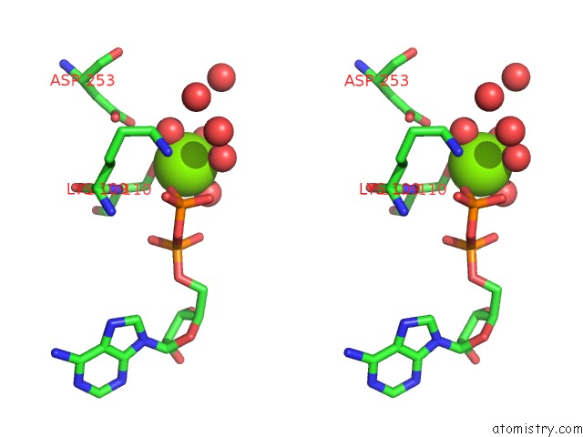
Stereo pair view
Mono view
Stereo pair view
A full contact list of Magnesium with other atoms in the Mg binding
site number 1 of Crystal Structure of Auto-Inhibited Kinesin-3 KIF13B within 5.0Å range:
|
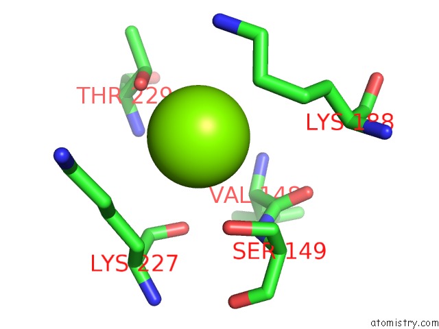
Magnesium binding site 2 out of 2 in 6a20
Go back to
Magnesium binding site 2 out
of 2 in the Crystal Structure of Auto-Inhibited Kinesin-3 KIF13B
Mono view
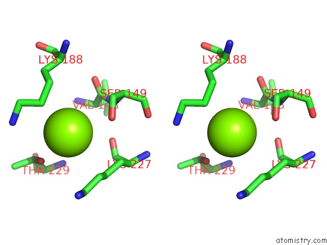
Stereo pair view
Mono view
Stereo pair view
A full contact list of Magnesium with other atoms in the Mg binding
site number 2 of Crystal Structure of Auto-Inhibited Kinesin-3 KIF13B within 5.0Å range:
|
Reference:
J.Q.Ren,
S.Wang,
H.Chen,
W.J.Wang,
L.Huo,
W.Feng.
Coiled-Coil 1-Mediated Fastening of the Neck and Motor Domains For Kinesin-3 Autoinhibition. Proc. Natl. Acad. Sci. V. 115 11933 2018U.S.A..
ISSN: ESSN 1091-6490
PubMed: 30463954
DOI: 10.1073/PNAS.1811209115
Page generated: Mon Sep 30 18:55:51 2024
ISSN: ESSN 1091-6490
PubMed: 30463954
DOI: 10.1073/PNAS.1811209115
Last articles
Zn in 9MJ5Zn in 9HNW
Zn in 9G0L
Zn in 9FNE
Zn in 9DZN
Zn in 9E0I
Zn in 9D32
Zn in 9DAK
Zn in 8ZXC
Zn in 8ZUF