Magnesium »
PDB 6a1m-6aad »
6a5s »
Magnesium in PDB 6a5s: Structure of 14-3-3 Gamma in Complex with Tfeb 14-3-3 Binding Motif
Protein crystallography data
The structure of Structure of 14-3-3 Gamma in Complex with Tfeb 14-3-3 Binding Motif, PDB code: 6a5s
was solved by
Y.Xu,
J.Q.Ren,
W.Feng,
with X-Ray Crystallography technique. A brief refinement statistics is given in the table below:
| Resolution Low / High (Å) | 44.04 / 2.10 |
| Space group | P 1 21 1 |
| Cell size a, b, c (Å), α, β, γ (°) | 88.088, 82.229, 88.092, 90.00, 90.11, 90.00 |
| R / Rfree (%) | 18 / 22.7 |
Other elements in 6a5s:
The structure of Structure of 14-3-3 Gamma in Complex with Tfeb 14-3-3 Binding Motif also contains other interesting chemical elements:
| Sodium | (Na) | 1 atom |
Magnesium Binding Sites:
The binding sites of Magnesium atom in the Structure of 14-3-3 Gamma in Complex with Tfeb 14-3-3 Binding Motif
(pdb code 6a5s). This binding sites where shown within
5.0 Angstroms radius around Magnesium atom.
In total only one binding site of Magnesium was determined in the Structure of 14-3-3 Gamma in Complex with Tfeb 14-3-3 Binding Motif, PDB code: 6a5s:
In total only one binding site of Magnesium was determined in the Structure of 14-3-3 Gamma in Complex with Tfeb 14-3-3 Binding Motif, PDB code: 6a5s:
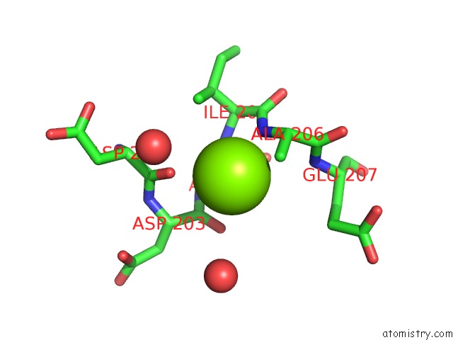
Magnesium binding site 1 out of 1 in 6a5s
Go back to
Magnesium binding site 1 out
of 1 in the Structure of 14-3-3 Gamma in Complex with Tfeb 14-3-3 Binding Motif
Mono view

Stereo pair view
Mono view
Stereo pair view
A full contact list of Magnesium with other atoms in the Mg binding
site number 1 of Structure of 14-3-3 Gamma in Complex with Tfeb 14-3-3 Binding Motif within 5.0Å range:
|
Reference:
Y.Xu,
J.Ren,
X.He,
H.Chen,
T.Wei,
W.Feng.
Ywha/14-3-3 Proteins Recognize Phosphorylated Tfeb By A Noncanonical Mode For Controlling Tfeb Cytoplasmic Localization. Autophagy V. 15 1017 2019.
ISSN: ESSN 1554-8635
PubMed: 30653408
DOI: 10.1080/15548627.2019.1569928
Page generated: Mon Sep 30 18:59:38 2024
ISSN: ESSN 1554-8635
PubMed: 30653408
DOI: 10.1080/15548627.2019.1569928
Last articles
Ca in 5UG7Ca in 5UII
Ca in 5UGO
Ca in 5UFQ
Ca in 5UE4
Ca in 5UE5
Ca in 5UFE
Ca in 5UE3
Ca in 5UE1
Ca in 5UE2