Magnesium »
PDB 6a1m-6aad »
6a6m »
Magnesium in PDB 6a6m: Crystal Structure of An Outward-Open Nucleotide-Bound State of the Eukaryotic Abc Multidrug Transporter CMABCB1
Protein crystallography data
The structure of Crystal Structure of An Outward-Open Nucleotide-Bound State of the Eukaryotic Abc Multidrug Transporter CMABCB1, PDB code: 6a6m
was solved by
H.Kato,
T.Nakatsu,
A.Kodan,
with X-Ray Crystallography technique. A brief refinement statistics is given in the table below:
| Resolution Low / High (Å) | 43.62 / 1.90 |
| Space group | P 41 3 2 |
| Cell size a, b, c (Å), α, β, γ (°) | 174.340, 174.340, 174.340, 90.00, 90.00, 90.00 |
| R / Rfree (%) | 16.5 / 20.8 |
Magnesium Binding Sites:
The binding sites of Magnesium atom in the Crystal Structure of An Outward-Open Nucleotide-Bound State of the Eukaryotic Abc Multidrug Transporter CMABCB1
(pdb code 6a6m). This binding sites where shown within
5.0 Angstroms radius around Magnesium atom.
In total only one binding site of Magnesium was determined in the Crystal Structure of An Outward-Open Nucleotide-Bound State of the Eukaryotic Abc Multidrug Transporter CMABCB1, PDB code: 6a6m:
In total only one binding site of Magnesium was determined in the Crystal Structure of An Outward-Open Nucleotide-Bound State of the Eukaryotic Abc Multidrug Transporter CMABCB1, PDB code: 6a6m:
Magnesium binding site 1 out of 1 in 6a6m
Go back to
Magnesium binding site 1 out
of 1 in the Crystal Structure of An Outward-Open Nucleotide-Bound State of the Eukaryotic Abc Multidrug Transporter CMABCB1
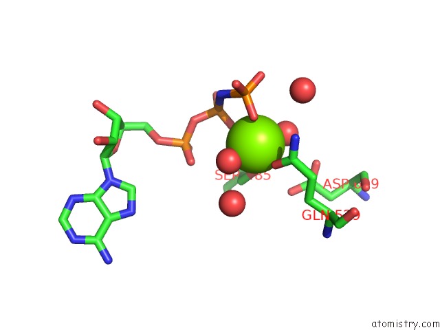
Mono view
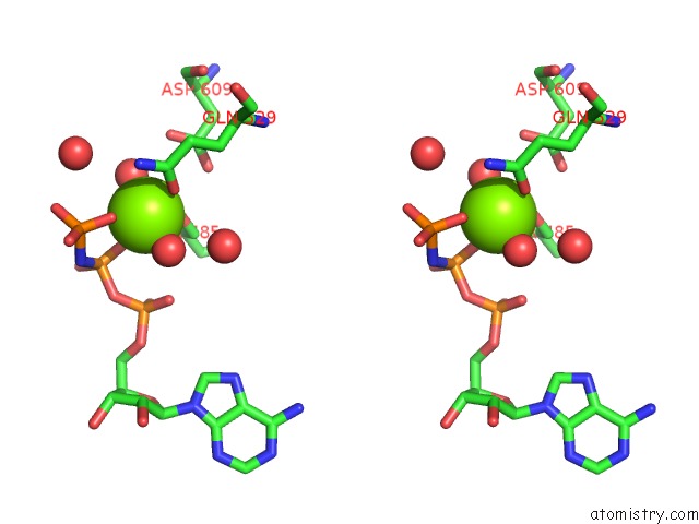
Stereo pair view
Mono view
Stereo pair view
A full contact list of Magnesium with other atoms in the Mg binding
site number 1 of Crystal Structure of An Outward-Open Nucleotide-Bound State of the Eukaryotic Abc Multidrug Transporter CMABCB1 within 5.0Å range:
|
Reference:
A.Kodan,
T.Yamaguchi,
T.Nakatsu,
K.Matsuoka,
Y.Kimura,
K.Ueda,
H.Kato.
Inward- and Outward-Facing X-Ray Crystal Structures of Homodimeric P-Glycoprotein CMABCB1. Nat Commun V. 10 88 2019.
ISSN: ESSN 2041-1723
PubMed: 30622258
DOI: 10.1038/S41467-018-08007-X
Page generated: Mon Sep 30 19:00:19 2024
ISSN: ESSN 2041-1723
PubMed: 30622258
DOI: 10.1038/S41467-018-08007-X
Last articles
Zn in 9MJ5Zn in 9HNW
Zn in 9G0L
Zn in 9FNE
Zn in 9DZN
Zn in 9E0I
Zn in 9D32
Zn in 9DAK
Zn in 8ZXC
Zn in 8ZUF