Magnesium »
PDB 6ako-6asu »
6asg »
Magnesium in PDB 6asg: Crystal Structure of Thermus Thermophilus Rna Polymerase Core Enzyme
Enzymatic activity of Crystal Structure of Thermus Thermophilus Rna Polymerase Core Enzyme
All present enzymatic activity of Crystal Structure of Thermus Thermophilus Rna Polymerase Core Enzyme:
2.7.7.6;
2.7.7.6;
Protein crystallography data
The structure of Crystal Structure of Thermus Thermophilus Rna Polymerase Core Enzyme, PDB code: 6asg
was solved by
Y.Liu,
W.Lin,
R.Ying,
R.H.Ebright,
with X-Ray Crystallography technique. A brief refinement statistics is given in the table below:
| Resolution Low / High (Å) | 42.49 / 3.80 |
| Space group | H 3 |
| Cell size a, b, c (Å), α, β, γ (°) | 280.694, 280.694, 184.964, 90.00, 90.00, 120.00 |
| R / Rfree (%) | 22.9 / 27.8 |
Other elements in 6asg:
The structure of Crystal Structure of Thermus Thermophilus Rna Polymerase Core Enzyme also contains other interesting chemical elements:
| Zinc | (Zn) | 2 atoms |
Magnesium Binding Sites:
The binding sites of Magnesium atom in the Crystal Structure of Thermus Thermophilus Rna Polymerase Core Enzyme
(pdb code 6asg). This binding sites where shown within
5.0 Angstroms radius around Magnesium atom.
In total only one binding site of Magnesium was determined in the Crystal Structure of Thermus Thermophilus Rna Polymerase Core Enzyme, PDB code: 6asg:
In total only one binding site of Magnesium was determined in the Crystal Structure of Thermus Thermophilus Rna Polymerase Core Enzyme, PDB code: 6asg:
Magnesium binding site 1 out of 1 in 6asg
Go back to
Magnesium binding site 1 out
of 1 in the Crystal Structure of Thermus Thermophilus Rna Polymerase Core Enzyme
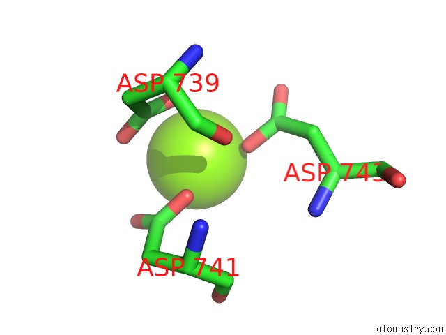
Mono view
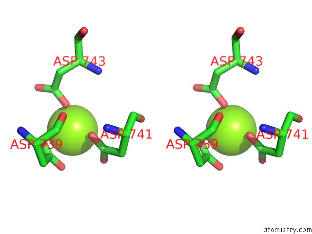
Stereo pair view
Mono view
Stereo pair view
A full contact list of Magnesium with other atoms in the Mg binding
site number 1 of Crystal Structure of Thermus Thermophilus Rna Polymerase Core Enzyme within 5.0Å range:
|
Reference:
W.Lin,
K.Das,
D.Degen,
A.Mazumder,
D.Duchi,
D.Wang,
Y.W.Ebright,
R.Y.Ebright,
E.Sineva,
M.Gigliotti,
A.Srivastava,
S.Mandal,
Y.Jiang,
Y.Liu,
R.Yin,
Z.Zhang,
E.T.Eng,
D.Thomas,
S.Donadio,
H.Zhang,
C.Zhang,
A.N.Kapanidis,
R.H.Ebright.
Structural Basis of Transcription Inhibition By Fidaxomicin (Lipiarmycin A3). Mol. Cell V. 70 60 2018.
ISSN: ISSN 1097-4164
PubMed: 29606590
DOI: 10.1016/J.MOLCEL.2018.02.026
Page generated: Mon Sep 30 19:20:35 2024
ISSN: ISSN 1097-4164
PubMed: 29606590
DOI: 10.1016/J.MOLCEL.2018.02.026
Last articles
Zn in 9J0NZn in 9J0O
Zn in 9J0P
Zn in 9FJX
Zn in 9EKB
Zn in 9C0F
Zn in 9CAH
Zn in 9CH0
Zn in 9CH3
Zn in 9CH1