Magnesium »
PDB 6asw-6b4k »
6ayu »
Magnesium in PDB 6ayu: Crystal Structure of Fructose-1,6-Bisphosphatase T84S From Mycobacterium Tuberculosis
Enzymatic activity of Crystal Structure of Fructose-1,6-Bisphosphatase T84S From Mycobacterium Tuberculosis
All present enzymatic activity of Crystal Structure of Fructose-1,6-Bisphosphatase T84S From Mycobacterium Tuberculosis:
3.1.3.11;
3.1.3.11;
Protein crystallography data
The structure of Crystal Structure of Fructose-1,6-Bisphosphatase T84S From Mycobacterium Tuberculosis, PDB code: 6ayu
was solved by
C.Abad-Zapatero,
N.Wolf,
H.J.Gutka,
F.Movahedzadeh,
with X-Ray Crystallography technique. A brief refinement statistics is given in the table below:
| Resolution Low / High (Å) | 38.50 / 2.20 |
| Space group | P 61 2 2 |
| Cell size a, b, c (Å), α, β, γ (°) | 130.890, 130.890, 142.848, 90.00, 90.00, 120.00 |
| R / Rfree (%) | 20.4 / 25.2 |
Magnesium Binding Sites:
The binding sites of Magnesium atom in the Crystal Structure of Fructose-1,6-Bisphosphatase T84S From Mycobacterium Tuberculosis
(pdb code 6ayu). This binding sites where shown within
5.0 Angstroms radius around Magnesium atom.
In total 3 binding sites of Magnesium where determined in the Crystal Structure of Fructose-1,6-Bisphosphatase T84S From Mycobacterium Tuberculosis, PDB code: 6ayu:
Jump to Magnesium binding site number: 1; 2; 3;
In total 3 binding sites of Magnesium where determined in the Crystal Structure of Fructose-1,6-Bisphosphatase T84S From Mycobacterium Tuberculosis, PDB code: 6ayu:
Jump to Magnesium binding site number: 1; 2; 3;
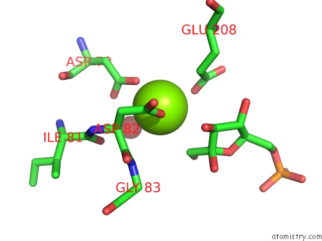
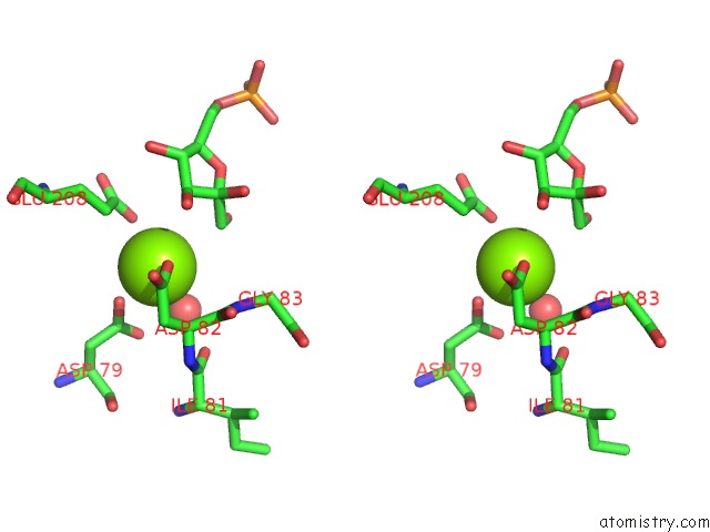
Magnesium binding site 1 out of 3 in 6ayu
Go back to
Magnesium binding site 1 out
of 3 in the Crystal Structure of Fructose-1,6-Bisphosphatase T84S From Mycobacterium Tuberculosis
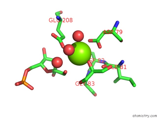
Mono view
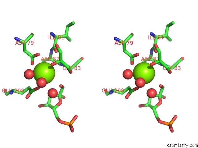
Stereo pair view
Mono view
Stereo pair view
A full contact list of Magnesium with other atoms in the Mg binding
site number 1 of Crystal Structure of Fructose-1,6-Bisphosphatase T84S From Mycobacterium Tuberculosis within 5.0Å range:
|
Magnesium binding site 2 out of 3 in 6ayu
Go back to
Magnesium binding site 2 out
of 3 in the Crystal Structure of Fructose-1,6-Bisphosphatase T84S From Mycobacterium Tuberculosis
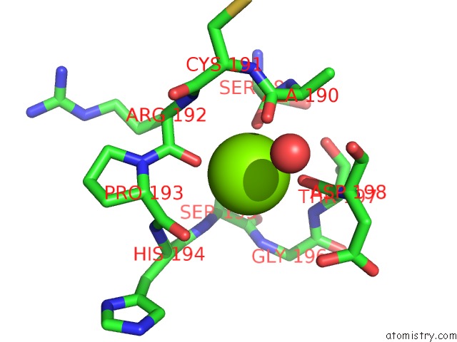
Mono view
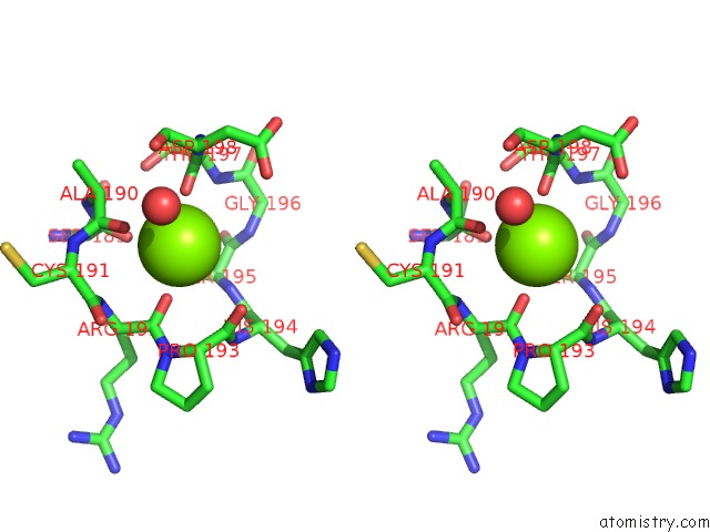
Stereo pair view
Mono view
Stereo pair view
A full contact list of Magnesium with other atoms in the Mg binding
site number 2 of Crystal Structure of Fructose-1,6-Bisphosphatase T84S From Mycobacterium Tuberculosis within 5.0Å range:
|
Magnesium binding site 3 out of 3 in 6ayu
Go back to
Magnesium binding site 3 out
of 3 in the Crystal Structure of Fructose-1,6-Bisphosphatase T84S From Mycobacterium Tuberculosis
Mono view
Stereo pair view
Mono view
Stereo pair view
A full contact list of Magnesium with other atoms in the Mg binding
site number 3 of Crystal Structure of Fructose-1,6-Bisphosphatase T84S From Mycobacterium Tuberculosis within 5.0Å range:
|
Reference:
N.M.Wolf,
H.J.Gutka,
F.Movahedzadeh,
C.Abad-Zapatero.
Structures of the Mycobacterium Tuberculosis Glpx Protein (Class II Fructose-1,6-Bisphosphatase): Implications For the Active Oligomeric State, Catalytic Mechanism and Citrate Inhibition. Acta Crystallogr D Struct V. 74 321 2018BIOL.
ISSN: ISSN 2059-7983
PubMed: 29652259
DOI: 10.1107/S2059798318002838
Page generated: Mon Sep 30 19:24:56 2024
ISSN: ISSN 2059-7983
PubMed: 29652259
DOI: 10.1107/S2059798318002838
Last articles
Zn in 9J0NZn in 9J0O
Zn in 9J0P
Zn in 9FJX
Zn in 9EKB
Zn in 9C0F
Zn in 9CAH
Zn in 9CH0
Zn in 9CH3
Zn in 9CH1