Magnesium »
PDB 6asw-6b4k »
6b09 »
Magnesium in PDB 6b09: Crystal Structure of HSNUDT16 in Complex with Diadpr (Soaked)
Enzymatic activity of Crystal Structure of HSNUDT16 in Complex with Diadpr (Soaked)
All present enzymatic activity of Crystal Structure of HSNUDT16 in Complex with Diadpr (Soaked):
3.6.1.62; 3.6.1.64;
3.6.1.62; 3.6.1.64;
Protein crystallography data
The structure of Crystal Structure of HSNUDT16 in Complex with Diadpr (Soaked), PDB code: 6b09
was solved by
P.Thirawatananond,
S.B.Gabelli,
with X-Ray Crystallography technique. A brief refinement statistics is given in the table below:
| Resolution Low / High (Å) | 37.62 / 3.20 |
| Space group | C 1 2 1 |
| Cell size a, b, c (Å), α, β, γ (°) | 113.395, 47.063, 75.792, 90.00, 108.74, 90.00 |
| R / Rfree (%) | 25.6 / 27.9 |
Other elements in 6b09:
The structure of Crystal Structure of HSNUDT16 in Complex with Diadpr (Soaked) also contains other interesting chemical elements:
| Chlorine | (Cl) | 1 atom |
| Sodium | (Na) | 2 atoms |
Magnesium Binding Sites:
The binding sites of Magnesium atom in the Crystal Structure of HSNUDT16 in Complex with Diadpr (Soaked)
(pdb code 6b09). This binding sites where shown within
5.0 Angstroms radius around Magnesium atom.
In total 4 binding sites of Magnesium where determined in the Crystal Structure of HSNUDT16 in Complex with Diadpr (Soaked), PDB code: 6b09:
Jump to Magnesium binding site number: 1; 2; 3; 4;
In total 4 binding sites of Magnesium where determined in the Crystal Structure of HSNUDT16 in Complex with Diadpr (Soaked), PDB code: 6b09:
Jump to Magnesium binding site number: 1; 2; 3; 4;
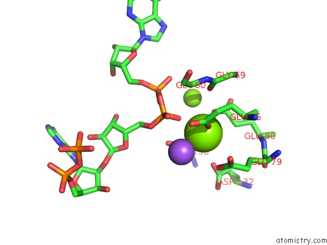
Magnesium binding site 1 out of 4 in 6b09
Go back to
Magnesium binding site 1 out
of 4 in the Crystal Structure of HSNUDT16 in Complex with Diadpr (Soaked)

Mono view

Stereo pair view
Mono view
Stereo pair view
A full contact list of Magnesium with other atoms in the Mg binding
site number 1 of Crystal Structure of HSNUDT16 in Complex with Diadpr (Soaked) within 5.0Å range:
|
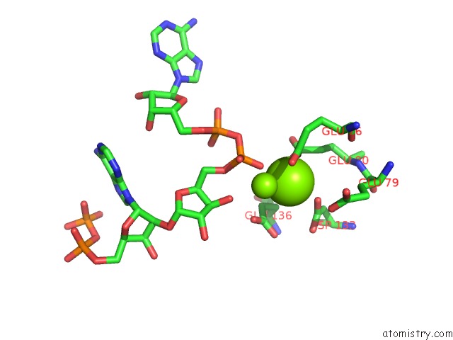
Magnesium binding site 2 out of 4 in 6b09
Go back to
Magnesium binding site 2 out
of 4 in the Crystal Structure of HSNUDT16 in Complex with Diadpr (Soaked)
Mono view

Stereo pair view
Mono view
Stereo pair view
A full contact list of Magnesium with other atoms in the Mg binding
site number 2 of Crystal Structure of HSNUDT16 in Complex with Diadpr (Soaked) within 5.0Å range:
|
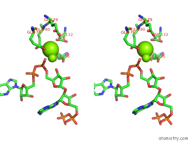
Magnesium binding site 3 out of 4 in 6b09
Go back to
Magnesium binding site 3 out
of 4 in the Crystal Structure of HSNUDT16 in Complex with Diadpr (Soaked)

Mono view

Stereo pair view
Mono view
Stereo pair view
A full contact list of Magnesium with other atoms in the Mg binding
site number 3 of Crystal Structure of HSNUDT16 in Complex with Diadpr (Soaked) within 5.0Å range:
|
Magnesium binding site 4 out of 4 in 6b09
Go back to
Magnesium binding site 4 out
of 4 in the Crystal Structure of HSNUDT16 in Complex with Diadpr (Soaked)
Mono view
Stereo pair view
Mono view
Stereo pair view
A full contact list of Magnesium with other atoms in the Mg binding
site number 4 of Crystal Structure of HSNUDT16 in Complex with Diadpr (Soaked) within 5.0Å range:
|
Reference:
P.Thirawatananond,
R.L.Mcpherson,
J.Malhi,
S.Nathan,
M.J.Lambrecht,
M.Brichacek,
P.J.Hergenrother,
A.K.L.Leung,
S.B.Gabelli.
Structural Analyses of NUDT16-Adp-Ribose Complexes Direct Rational Design of Mutants with Improved Processing of Poly(Adp-Ribosyl)Ated Proteins. Sci Rep V. 9 5940 2019.
ISSN: ESSN 2045-2322
PubMed: 30976021
DOI: 10.1038/S41598-019-39491-W
Page generated: Mon Sep 30 19:26:51 2024
ISSN: ESSN 2045-2322
PubMed: 30976021
DOI: 10.1038/S41598-019-39491-W
Last articles
Zn in 9J0NZn in 9J0O
Zn in 9J0P
Zn in 9FJX
Zn in 9EKB
Zn in 9C0F
Zn in 9CAH
Zn in 9CH0
Zn in 9CH3
Zn in 9CH1