Magnesium »
PDB 6b4o-6bcb »
6b6u »
Magnesium in PDB 6b6u: Pyruvate Kinase M2 Mutant - S437Y
Enzymatic activity of Pyruvate Kinase M2 Mutant - S437Y
All present enzymatic activity of Pyruvate Kinase M2 Mutant - S437Y:
2.7.1.40;
2.7.1.40;
Protein crystallography data
The structure of Pyruvate Kinase M2 Mutant - S437Y, PDB code: 6b6u
was solved by
D.Srivastava,
M.Dey,
with X-Ray Crystallography technique. A brief refinement statistics is given in the table below:
| Resolution Low / High (Å) | 28.99 / 1.35 |
| Space group | C 1 2 1 |
| Cell size a, b, c (Å), α, β, γ (°) | 110.350, 93.610, 109.360, 90.00, 95.64, 90.00 |
| R / Rfree (%) | 13 / 14.9 |
Other elements in 6b6u:
The structure of Pyruvate Kinase M2 Mutant - S437Y also contains other interesting chemical elements:
| Potassium | (K) | 2 atoms |
| Chlorine | (Cl) | 2 atoms |
Magnesium Binding Sites:
The binding sites of Magnesium atom in the Pyruvate Kinase M2 Mutant - S437Y
(pdb code 6b6u). This binding sites where shown within
5.0 Angstroms radius around Magnesium atom.
In total 2 binding sites of Magnesium where determined in the Pyruvate Kinase M2 Mutant - S437Y, PDB code: 6b6u:
Jump to Magnesium binding site number: 1; 2;
In total 2 binding sites of Magnesium where determined in the Pyruvate Kinase M2 Mutant - S437Y, PDB code: 6b6u:
Jump to Magnesium binding site number: 1; 2;
Magnesium binding site 1 out of 2 in 6b6u
Go back to
Magnesium binding site 1 out
of 2 in the Pyruvate Kinase M2 Mutant - S437Y
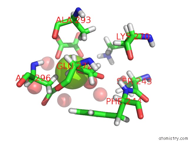
Mono view
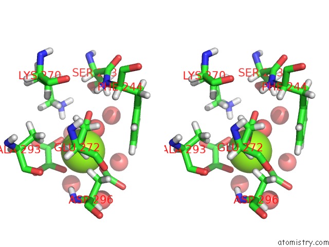
Stereo pair view
Mono view
Stereo pair view
A full contact list of Magnesium with other atoms in the Mg binding
site number 1 of Pyruvate Kinase M2 Mutant - S437Y within 5.0Å range:
|
Magnesium binding site 2 out of 2 in 6b6u
Go back to
Magnesium binding site 2 out
of 2 in the Pyruvate Kinase M2 Mutant - S437Y
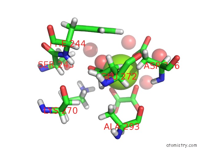
Mono view
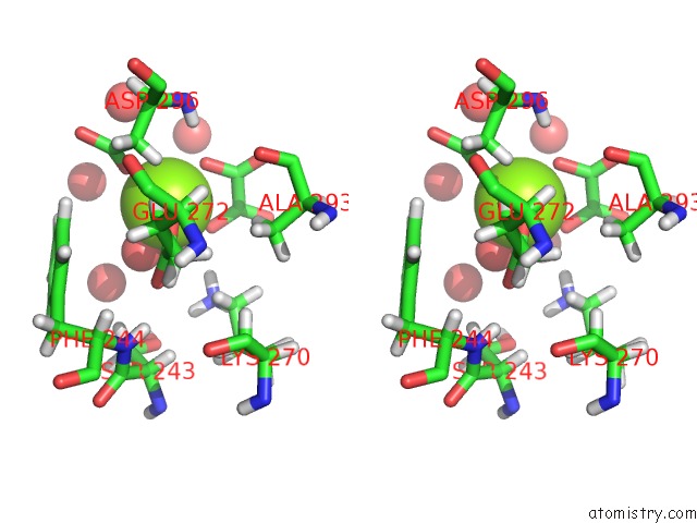
Stereo pair view
Mono view
Stereo pair view
A full contact list of Magnesium with other atoms in the Mg binding
site number 2 of Pyruvate Kinase M2 Mutant - S437Y within 5.0Å range:
|
Reference:
D.Srivastava,
M.Razzaghi,
M.T.Henzl,
M.Dey.
Structural Investigation of A Dimeric Variant of Pyruvate Kinase Muscle Isoform 2. Biochemistry V. 56 6517 2017.
ISSN: ISSN 1520-4995
PubMed: 29182273
DOI: 10.1021/ACS.BIOCHEM.7B01013
Page generated: Mon Sep 30 19:35:56 2024
ISSN: ISSN 1520-4995
PubMed: 29182273
DOI: 10.1021/ACS.BIOCHEM.7B01013
Last articles
Zn in 9J0NZn in 9J0O
Zn in 9J0P
Zn in 9FJX
Zn in 9EKB
Zn in 9C0F
Zn in 9CAH
Zn in 9CH0
Zn in 9CH3
Zn in 9CH1