Magnesium »
PDB 6bd1-6br7 »
6bjr »
Magnesium in PDB 6bjr: Crystal Structure of Prothrombin Mutant S101C/A470C
Enzymatic activity of Crystal Structure of Prothrombin Mutant S101C/A470C
All present enzymatic activity of Crystal Structure of Prothrombin Mutant S101C/A470C:
3.4.21.5;
3.4.21.5;
Protein crystallography data
The structure of Crystal Structure of Prothrombin Mutant S101C/A470C, PDB code: 6bjr
was solved by
M.Chinnaraj,
Z.Chen,
L.Pelc,
Z.Grese,
D.Bystranowska,
E.Di Cera,
N.Pozzi,
with X-Ray Crystallography technique. A brief refinement statistics is given in the table below:
| Resolution Low / High (Å) | 40.00 / 6.00 |
| Space group | C 2 2 21 |
| Cell size a, b, c (Å), α, β, γ (°) | 114.126, 124.175, 157.093, 90.00, 90.00, 90.00 |
| R / Rfree (%) | 23.7 / 29.3 |
Magnesium Binding Sites:
The binding sites of Magnesium atom in the Crystal Structure of Prothrombin Mutant S101C/A470C
(pdb code 6bjr). This binding sites where shown within
5.0 Angstroms radius around Magnesium atom.
In total 6 binding sites of Magnesium where determined in the Crystal Structure of Prothrombin Mutant S101C/A470C, PDB code: 6bjr:
Jump to Magnesium binding site number: 1; 2; 3; 4; 5; 6;
In total 6 binding sites of Magnesium where determined in the Crystal Structure of Prothrombin Mutant S101C/A470C, PDB code: 6bjr:
Jump to Magnesium binding site number: 1; 2; 3; 4; 5; 6;
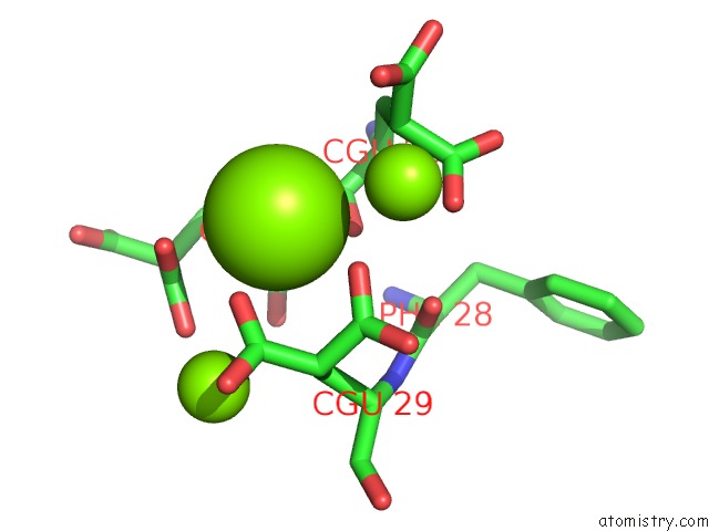
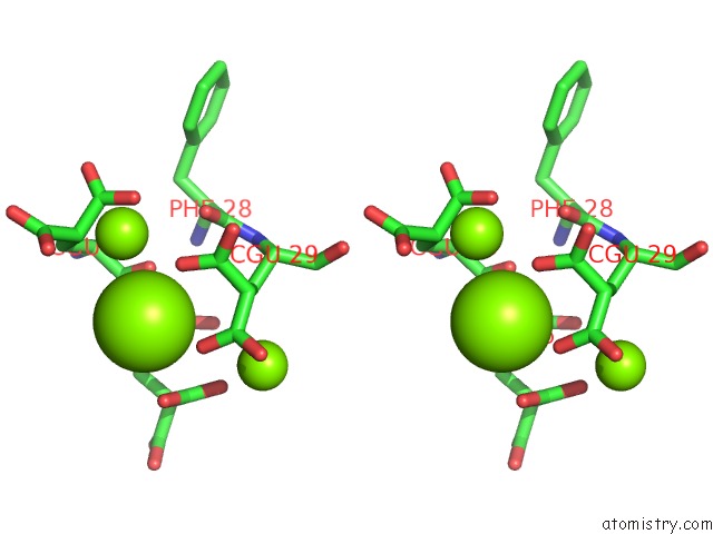
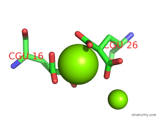
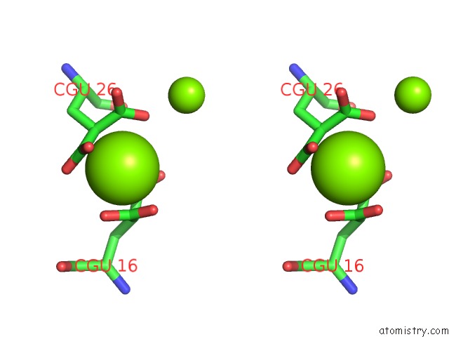
Magnesium binding site 1 out of 6 in 6bjr
Go back to
Magnesium binding site 1 out
of 6 in the Crystal Structure of Prothrombin Mutant S101C/A470C
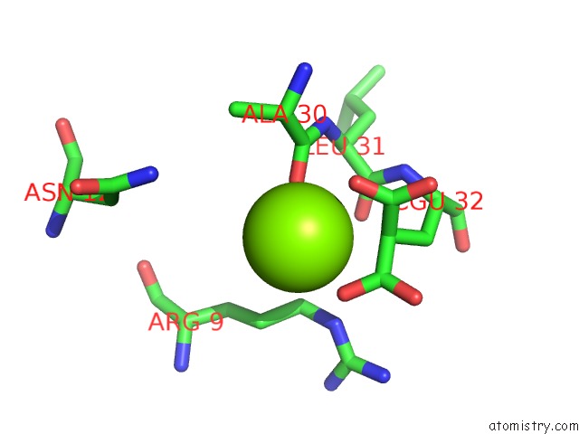
Mono view
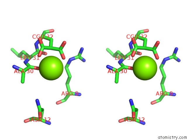
Stereo pair view
Mono view
Stereo pair view
A full contact list of Magnesium with other atoms in the Mg binding
site number 1 of Crystal Structure of Prothrombin Mutant S101C/A470C within 5.0Å range:
|
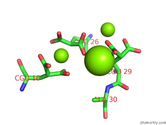
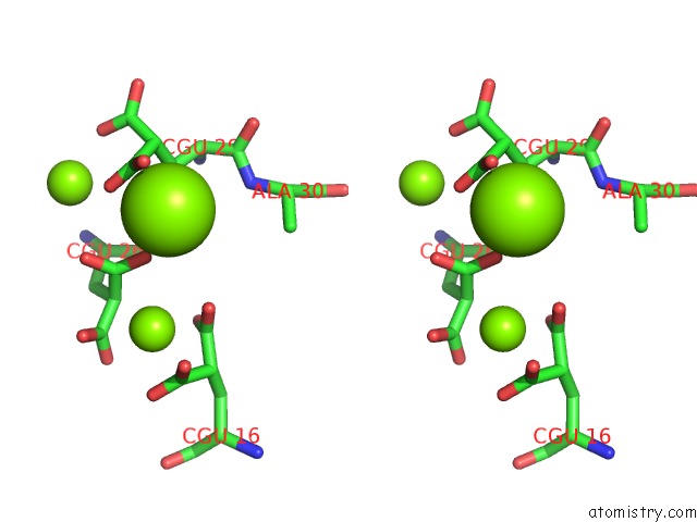
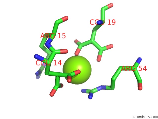
Magnesium binding site 2 out of 6 in 6bjr
Go back to
Magnesium binding site 2 out
of 6 in the Crystal Structure of Prothrombin Mutant S101C/A470C
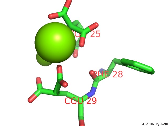
Mono view
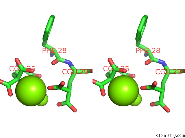
Stereo pair view
Mono view
Stereo pair view
A full contact list of Magnesium with other atoms in the Mg binding
site number 2 of Crystal Structure of Prothrombin Mutant S101C/A470C within 5.0Å range:
|
Magnesium binding site 3 out of 6 in 6bjr
Go back to
Magnesium binding site 3 out
of 6 in the Crystal Structure of Prothrombin Mutant S101C/A470C
Mono view
Stereo pair view
Mono view
Stereo pair view
A full contact list of Magnesium with other atoms in the Mg binding
site number 3 of Crystal Structure of Prothrombin Mutant S101C/A470C within 5.0Å range:
|
Magnesium binding site 4 out of 6 in 6bjr
Go back to
Magnesium binding site 4 out
of 6 in the Crystal Structure of Prothrombin Mutant S101C/A470C
Mono view
Stereo pair view
Mono view
Stereo pair view
A full contact list of Magnesium with other atoms in the Mg binding
site number 4 of Crystal Structure of Prothrombin Mutant S101C/A470C within 5.0Å range:
|
Magnesium binding site 5 out of 6 in 6bjr
Go back to
Magnesium binding site 5 out
of 6 in the Crystal Structure of Prothrombin Mutant S101C/A470C
Mono view
Stereo pair view
Mono view
Stereo pair view
A full contact list of Magnesium with other atoms in the Mg binding
site number 5 of Crystal Structure of Prothrombin Mutant S101C/A470C within 5.0Å range:
|
Magnesium binding site 6 out of 6 in 6bjr
Go back to
Magnesium binding site 6 out
of 6 in the Crystal Structure of Prothrombin Mutant S101C/A470C
Mono view

Stereo pair view
Mono view
Stereo pair view
A full contact list of Magnesium with other atoms in the Mg binding
site number 6 of Crystal Structure of Prothrombin Mutant S101C/A470C within 5.0Å range:
|
Reference:
M.Chinnaraj,
Z.Chen,
L.A.Pelc,
Z.Grese,
D.Bystranowska,
E.Di Cera,
N.Pozzi.
Structure of Prothrombin in the Closed Form Reveals New Details on the Mechanism of Activation. Sci Rep V. 8 2945 2018.
ISSN: ESSN 2045-2322
PubMed: 29440720
DOI: 10.1038/S41598-018-21304-1
Page generated: Wed Aug 13 02:38:55 2025
ISSN: ESSN 2045-2322
PubMed: 29440720
DOI: 10.1038/S41598-018-21304-1
Last articles
Mg in 6EE4Mg in 6EEC
Mg in 6EE8
Mg in 6EE6
Mg in 6EDW
Mg in 6EE3
Mg in 6EE1
Mg in 6EAC
Mg in 6ED2
Mg in 6EDT