Magnesium »
PDB 6br1-6c06 »
6brd »
Magnesium in PDB 6brd: Crystal Structure of Rifampin Monooxygenase From Streptomyces Venezuelae, Complexed with Rifampin and Fad
Protein crystallography data
The structure of Crystal Structure of Rifampin Monooxygenase From Streptomyces Venezuelae, Complexed with Rifampin and Fad, PDB code: 6brd
was solved by
G.Cox,
J.Kelso,
P.J.Stogios,
A.Savchenko,
W.F.Anderson,
G.D.Wright,
Centerfor Structural Genomics Of Infectious Diseases (Csgid),
with X-Ray Crystallography technique. A brief refinement statistics is given in the table below:
| Resolution Low / High (Å) | 33.42 / 3.32 |
| Space group | C 1 2 1 |
| Cell size a, b, c (Å), α, β, γ (°) | 202.026, 129.402, 74.342, 90.00, 105.45, 90.00 |
| R / Rfree (%) | 18.5 / 23.4 |
Other elements in 6brd:
The structure of Crystal Structure of Rifampin Monooxygenase From Streptomyces Venezuelae, Complexed with Rifampin and Fad also contains other interesting chemical elements:
| Chlorine | (Cl) | 10 atoms |
Magnesium Binding Sites:
The binding sites of Magnesium atom in the Crystal Structure of Rifampin Monooxygenase From Streptomyces Venezuelae, Complexed with Rifampin and Fad
(pdb code 6brd). This binding sites where shown within
5.0 Angstroms radius around Magnesium atom.
In total only one binding site of Magnesium was determined in the Crystal Structure of Rifampin Monooxygenase From Streptomyces Venezuelae, Complexed with Rifampin and Fad, PDB code: 6brd:
In total only one binding site of Magnesium was determined in the Crystal Structure of Rifampin Monooxygenase From Streptomyces Venezuelae, Complexed with Rifampin and Fad, PDB code: 6brd:
Magnesium binding site 1 out of 1 in 6brd
Go back to
Magnesium binding site 1 out
of 1 in the Crystal Structure of Rifampin Monooxygenase From Streptomyces Venezuelae, Complexed with Rifampin and Fad
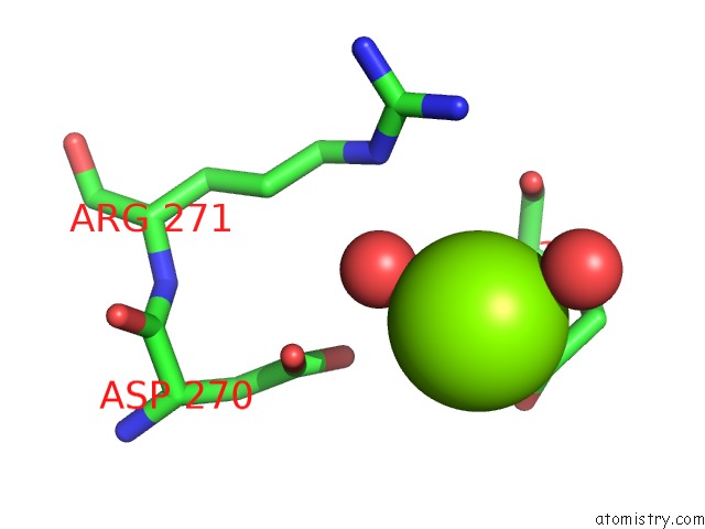
Mono view
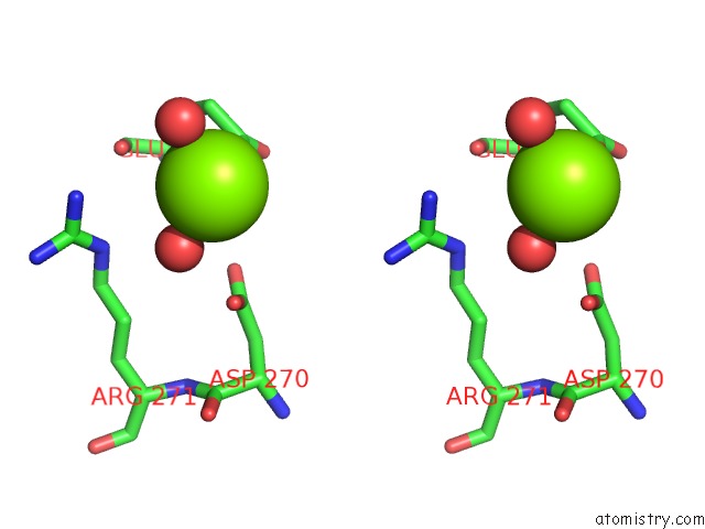
Stereo pair view
Mono view
Stereo pair view
A full contact list of Magnesium with other atoms in the Mg binding
site number 1 of Crystal Structure of Rifampin Monooxygenase From Streptomyces Venezuelae, Complexed with Rifampin and Fad within 5.0Å range:
|
Reference:
K.Koteva,
G.Cox,
J.K.Kelso,
M.D.Surette,
H.L.Zubyk,
L.Ejim,
P.Stogios,
A.Savchenko,
D.Sorensen,
G.D.Wright.
Rox, A Rifamycin Resistance Enzyme with An Unprecedented Mechanism of Action. Cell Chem Biol V. 25 403 2018.
ISSN: ESSN 2451-9448
PubMed: 29398560
DOI: 10.1016/J.CHEMBIOL.2018.01.009
Page generated: Mon Sep 30 19:57:40 2024
ISSN: ESSN 2451-9448
PubMed: 29398560
DOI: 10.1016/J.CHEMBIOL.2018.01.009
Last articles
Zn in 9J0NZn in 9J0O
Zn in 9J0P
Zn in 9FJX
Zn in 9EKB
Zn in 9C0F
Zn in 9CAH
Zn in 9CH0
Zn in 9CH3
Zn in 9CH1