Magnesium »
PDB 6br1-6c06 »
6bxj »
Magnesium in PDB 6bxj: Structure of A Single-Chain BETA3 Integrin
Protein crystallography data
The structure of Structure of A Single-Chain BETA3 Integrin, PDB code: 6bxj
was solved by
A.M.M.Thinn,
Z.Wang,
D.Zhou,
Y.Zhao,
B.R.Curtis,
J.Zhu,
with X-Ray Crystallography technique. A brief refinement statistics is given in the table below:
| Resolution Low / High (Å) | 47.29 / 2.09 |
| Space group | P 1 21 1 |
| Cell size a, b, c (Å), α, β, γ (°) | 55.460, 125.290, 70.020, 90.00, 112.95, 90.00 |
| R / Rfree (%) | 18.6 / 23.4 |
Magnesium Binding Sites:
The binding sites of Magnesium atom in the Structure of A Single-Chain BETA3 Integrin
(pdb code 6bxj). This binding sites where shown within
5.0 Angstroms radius around Magnesium atom.
In total 2 binding sites of Magnesium where determined in the Structure of A Single-Chain BETA3 Integrin, PDB code: 6bxj:
Jump to Magnesium binding site number: 1; 2;
In total 2 binding sites of Magnesium where determined in the Structure of A Single-Chain BETA3 Integrin, PDB code: 6bxj:
Jump to Magnesium binding site number: 1; 2;
Magnesium binding site 1 out of 2 in 6bxj
Go back to
Magnesium binding site 1 out
of 2 in the Structure of A Single-Chain BETA3 Integrin
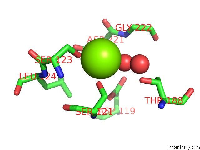
Mono view
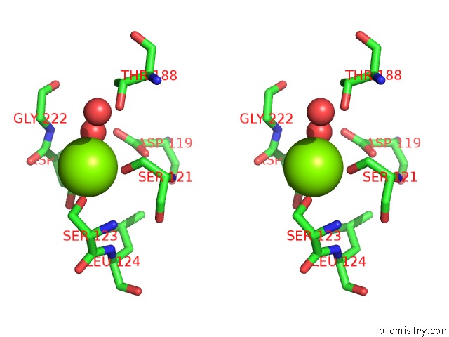
Stereo pair view
Mono view
Stereo pair view
A full contact list of Magnesium with other atoms in the Mg binding
site number 1 of Structure of A Single-Chain BETA3 Integrin within 5.0Å range:
|
Magnesium binding site 2 out of 2 in 6bxj
Go back to
Magnesium binding site 2 out
of 2 in the Structure of A Single-Chain BETA3 Integrin
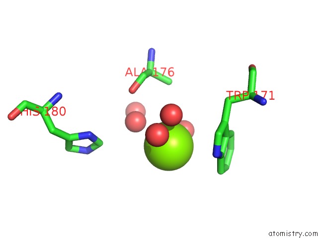
Mono view
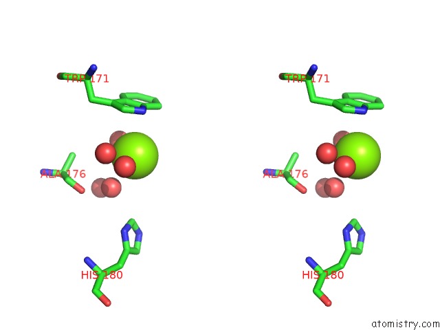
Stereo pair view
Mono view
Stereo pair view
A full contact list of Magnesium with other atoms in the Mg binding
site number 2 of Structure of A Single-Chain BETA3 Integrin within 5.0Å range:
|
Reference:
A.M.M.Thinn,
Z.Wang,
D.Zhou,
Y.Zhao,
B.R.Curtis,
J.Zhu.
Autonomous Conformational Regulation of BETA3INTEGRIN and the Conformation-Dependent Property of Hpa-1A Alloantibodies. Proc. Natl. Acad. Sci. V. 115 E9105 2018U.S.A..
ISSN: ESSN 1091-6490
PubMed: 30209215
DOI: 10.1073/PNAS.1806205115
Page generated: Mon Sep 30 20:02:54 2024
ISSN: ESSN 1091-6490
PubMed: 30209215
DOI: 10.1073/PNAS.1806205115
Last articles
Zn in 9JYWZn in 9IR4
Zn in 9IR3
Zn in 9GMX
Zn in 9GMW
Zn in 9JEJ
Zn in 9ERF
Zn in 9ERE
Zn in 9EGV
Zn in 9EGW