Magnesium »
PDB 6c0j-6c7j »
6c1x »
Magnesium in PDB 6c1x: Crystal Structure of Ketosteroid Isomerase D40N/D103N Mutant From Pseudomonas Putida (Pksi) Bound to 3,4-Dinitrophenol
Enzymatic activity of Crystal Structure of Ketosteroid Isomerase D40N/D103N Mutant From Pseudomonas Putida (Pksi) Bound to 3,4-Dinitrophenol
All present enzymatic activity of Crystal Structure of Ketosteroid Isomerase D40N/D103N Mutant From Pseudomonas Putida (Pksi) Bound to 3,4-Dinitrophenol:
5.3.3.1;
5.3.3.1;
Protein crystallography data
The structure of Crystal Structure of Ketosteroid Isomerase D40N/D103N Mutant From Pseudomonas Putida (Pksi) Bound to 3,4-Dinitrophenol, PDB code: 6c1x
was solved by
F.Yabukarski,
M.M.Pinney,
D.Herschlag,
with X-Ray Crystallography technique. A brief refinement statistics is given in the table below:
| Resolution Low / High (Å) | 36.50 / 1.05 |
| Space group | C 2 2 21 |
| Cell size a, b, c (Å), α, β, γ (°) | 36.386, 95.339, 72.999, 90.00, 90.00, 90.00 |
| R / Rfree (%) | 14.2 / 17.2 |
Magnesium Binding Sites:
The binding sites of Magnesium atom in the Crystal Structure of Ketosteroid Isomerase D40N/D103N Mutant From Pseudomonas Putida (Pksi) Bound to 3,4-Dinitrophenol
(pdb code 6c1x). This binding sites where shown within
5.0 Angstroms radius around Magnesium atom.
In total 2 binding sites of Magnesium where determined in the Crystal Structure of Ketosteroid Isomerase D40N/D103N Mutant From Pseudomonas Putida (Pksi) Bound to 3,4-Dinitrophenol, PDB code: 6c1x:
Jump to Magnesium binding site number: 1; 2;
In total 2 binding sites of Magnesium where determined in the Crystal Structure of Ketosteroid Isomerase D40N/D103N Mutant From Pseudomonas Putida (Pksi) Bound to 3,4-Dinitrophenol, PDB code: 6c1x:
Jump to Magnesium binding site number: 1; 2;
Magnesium binding site 1 out of 2 in 6c1x
Go back to
Magnesium binding site 1 out
of 2 in the Crystal Structure of Ketosteroid Isomerase D40N/D103N Mutant From Pseudomonas Putida (Pksi) Bound to 3,4-Dinitrophenol
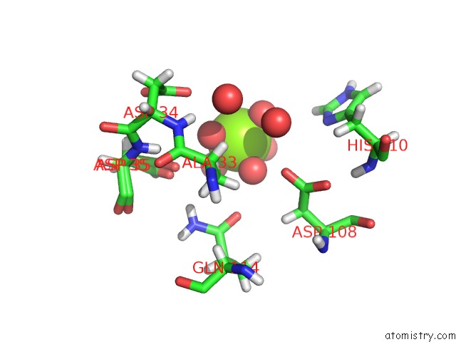
Mono view
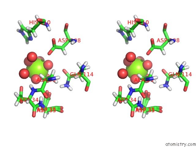
Stereo pair view
Mono view
Stereo pair view
A full contact list of Magnesium with other atoms in the Mg binding
site number 1 of Crystal Structure of Ketosteroid Isomerase D40N/D103N Mutant From Pseudomonas Putida (Pksi) Bound to 3,4-Dinitrophenol within 5.0Å range:
|
Magnesium binding site 2 out of 2 in 6c1x
Go back to
Magnesium binding site 2 out
of 2 in the Crystal Structure of Ketosteroid Isomerase D40N/D103N Mutant From Pseudomonas Putida (Pksi) Bound to 3,4-Dinitrophenol
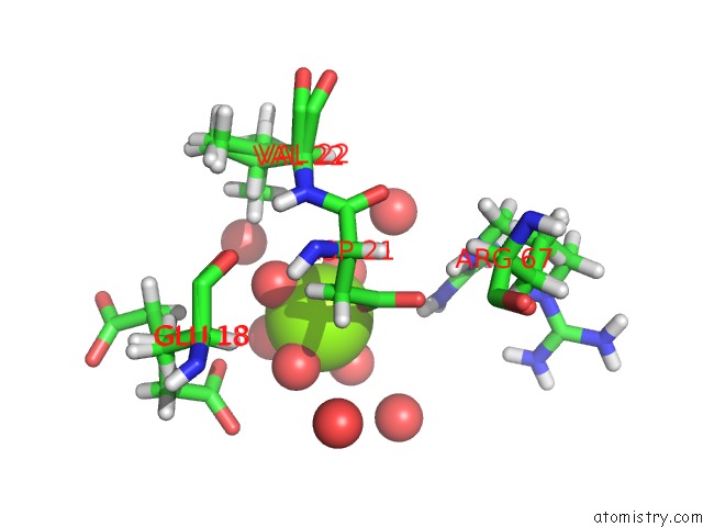
Mono view
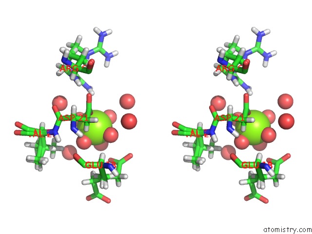
Stereo pair view
Mono view
Stereo pair view
A full contact list of Magnesium with other atoms in the Mg binding
site number 2 of Crystal Structure of Ketosteroid Isomerase D40N/D103N Mutant From Pseudomonas Putida (Pksi) Bound to 3,4-Dinitrophenol within 5.0Å range:
|
Reference:
M.M.Pinney,
A.Natarajan,
F.Yabukarski,
D.M.Sanchez,
F.Liu,
R.Liang,
T.Doukov,
J.P.Schwans,
T.J.Martinez,
D.Herschlag.
Structural Coupling Throughout the Active Site Hydrogen Bond Networks of Ketosteroid Isomerase and Photoactive Yellow Protein. J. Am. Chem. Soc. V. 140 9827 2018.
ISSN: ESSN 1520-5126
PubMed: 29990421
DOI: 10.1021/JACS.8B01596
Page generated: Mon Sep 30 20:05:54 2024
ISSN: ESSN 1520-5126
PubMed: 29990421
DOI: 10.1021/JACS.8B01596
Last articles
Zn in 9MJ5Zn in 9HNW
Zn in 9G0L
Zn in 9FNE
Zn in 9DZN
Zn in 9E0I
Zn in 9D32
Zn in 9DAK
Zn in 8ZXC
Zn in 8ZUF