Magnesium »
PDB 6cgf-6cq2 »
6ci0 »
Magnesium in PDB 6ci0: Catalytic Core Subunits (I and II) of Cytochrome C Oxidase From Rhodobacter Sphaeroides with E101A (II) Mutation
Enzymatic activity of Catalytic Core Subunits (I and II) of Cytochrome C Oxidase From Rhodobacter Sphaeroides with E101A (II) Mutation
All present enzymatic activity of Catalytic Core Subunits (I and II) of Cytochrome C Oxidase From Rhodobacter Sphaeroides with E101A (II) Mutation:
1.9.3.1;
1.9.3.1;
Protein crystallography data
The structure of Catalytic Core Subunits (I and II) of Cytochrome C Oxidase From Rhodobacter Sphaeroides with E101A (II) Mutation, PDB code: 6ci0
was solved by
J.Liu,
C.Hiser,
S.Ferguson-Miller,
with X-Ray Crystallography technique. A brief refinement statistics is given in the table below:
| Resolution Low / High (Å) | 42.09 / 2.40 |
| Space group | P 21 21 21 |
| Cell size a, b, c (Å), α, β, γ (°) | 124.916, 130.969, 177.788, 90.00, 90.00, 90.00 |
| R / Rfree (%) | 19.9 / 23.4 |
Other elements in 6ci0:
The structure of Catalytic Core Subunits (I and II) of Cytochrome C Oxidase From Rhodobacter Sphaeroides with E101A (II) Mutation also contains other interesting chemical elements:
| Potassium | (K) | 1 atom |
| Cadmium | (Cd) | 2 atoms |
| Iron | (Fe) | 4 atoms |
| Calcium | (Ca) | 2 atoms |
| Copper | (Cu) | 6 atoms |
Magnesium Binding Sites:
The binding sites of Magnesium atom in the Catalytic Core Subunits (I and II) of Cytochrome C Oxidase From Rhodobacter Sphaeroides with E101A (II) Mutation
(pdb code 6ci0). This binding sites where shown within
5.0 Angstroms radius around Magnesium atom.
In total 2 binding sites of Magnesium where determined in the Catalytic Core Subunits (I and II) of Cytochrome C Oxidase From Rhodobacter Sphaeroides with E101A (II) Mutation, PDB code: 6ci0:
Jump to Magnesium binding site number: 1; 2;
In total 2 binding sites of Magnesium where determined in the Catalytic Core Subunits (I and II) of Cytochrome C Oxidase From Rhodobacter Sphaeroides with E101A (II) Mutation, PDB code: 6ci0:
Jump to Magnesium binding site number: 1; 2;
Magnesium binding site 1 out of 2 in 6ci0
Go back to
Magnesium binding site 1 out
of 2 in the Catalytic Core Subunits (I and II) of Cytochrome C Oxidase From Rhodobacter Sphaeroides with E101A (II) Mutation
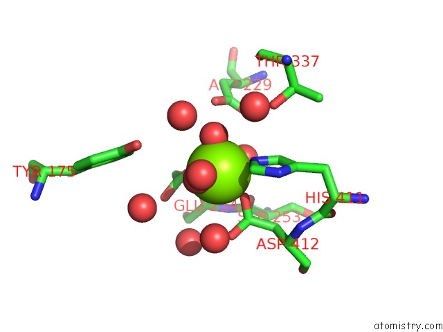
Mono view
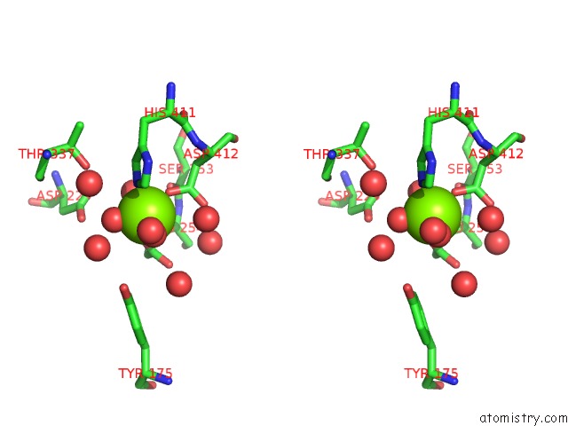
Stereo pair view
Mono view
Stereo pair view
A full contact list of Magnesium with other atoms in the Mg binding
site number 1 of Catalytic Core Subunits (I and II) of Cytochrome C Oxidase From Rhodobacter Sphaeroides with E101A (II) Mutation within 5.0Å range:
|
Magnesium binding site 2 out of 2 in 6ci0
Go back to
Magnesium binding site 2 out
of 2 in the Catalytic Core Subunits (I and II) of Cytochrome C Oxidase From Rhodobacter Sphaeroides with E101A (II) Mutation
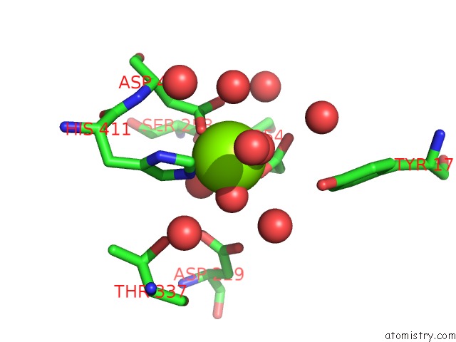
Mono view
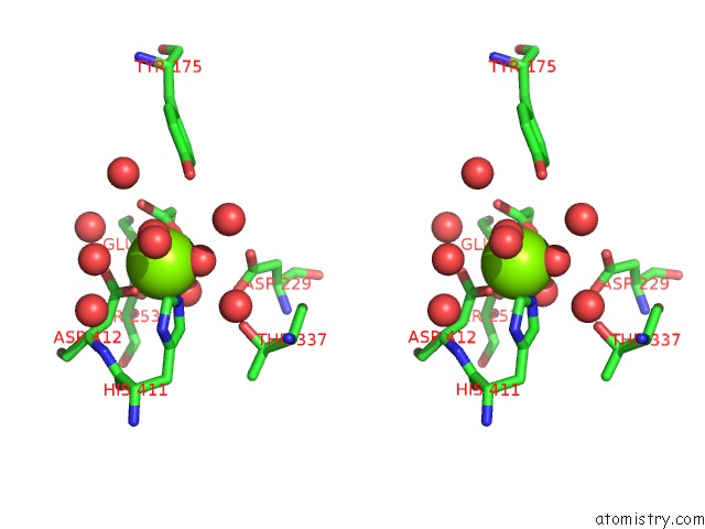
Stereo pair view
Mono view
Stereo pair view
A full contact list of Magnesium with other atoms in the Mg binding
site number 2 of Catalytic Core Subunits (I and II) of Cytochrome C Oxidase From Rhodobacter Sphaeroides with E101A (II) Mutation within 5.0Å range:
|
Reference:
C.Hiser,
J.Liu,
S.Ferguson-Miller.
The K-Path Entrance in Cytochrome C Oxidase Is Defined By Mutation of E101 and Controlled By An Adjacent Ligand Binding Domain. Biochim. Biophys. Acta V.1859 725 2018.
ISSN: ISSN 0006-3002
PubMed: 29626419
DOI: 10.1016/J.BBABIO.2018.03.017
Page generated: Wed Aug 13 04:31:00 2025
ISSN: ISSN 0006-3002
PubMed: 29626419
DOI: 10.1016/J.BBABIO.2018.03.017
Last articles
Mg in 6HNQMg in 6HOS
Mg in 6HNS
Mg in 6HN2
Mg in 6HMZ
Mg in 6HMU
Mg in 6HMT
Mg in 6HLR
Mg in 6HLQ
Mg in 6HKY